Posted on Leave a comment

Why GHK-Cu Is Everywhere in Skin and Tissue Science

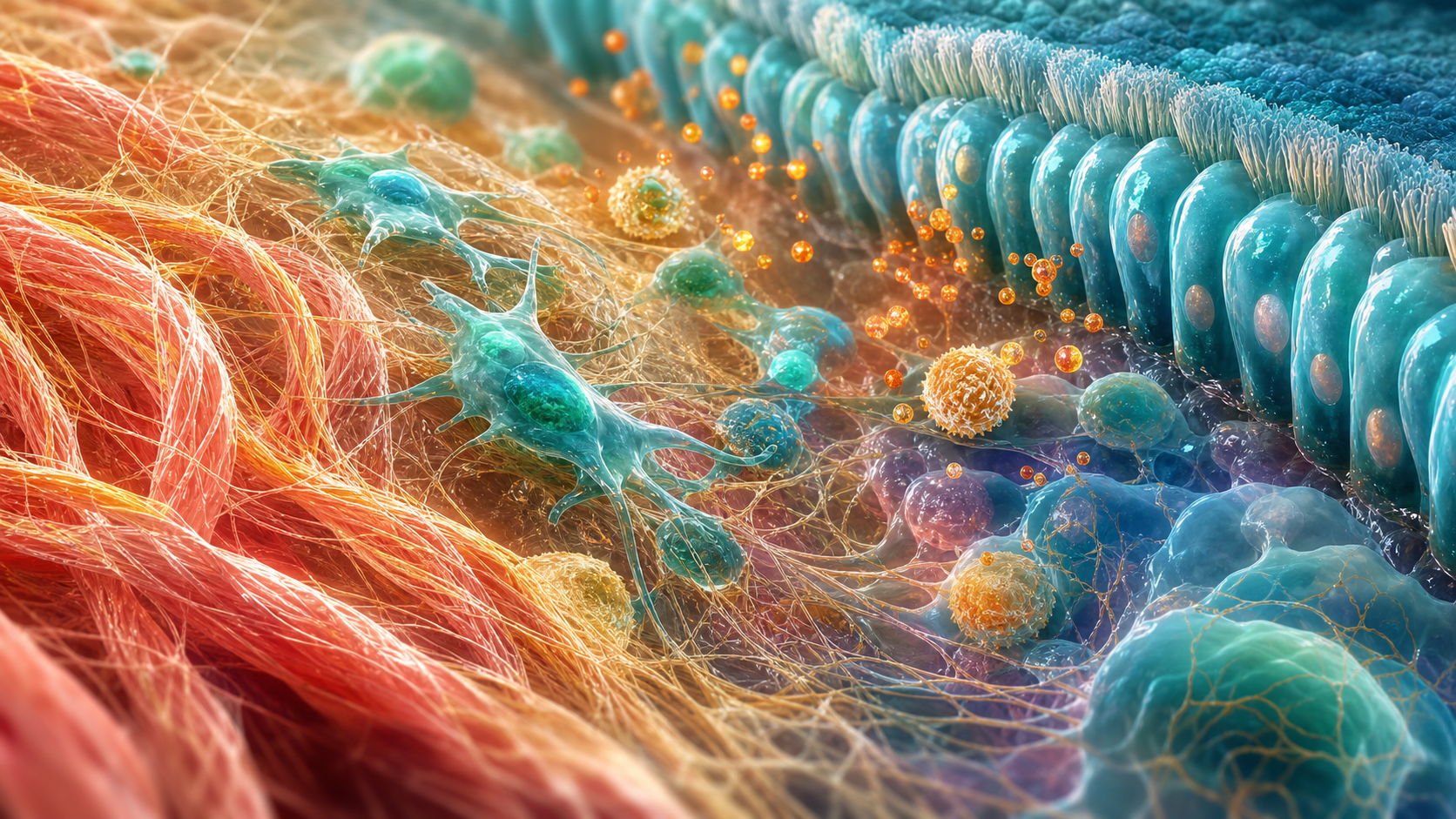
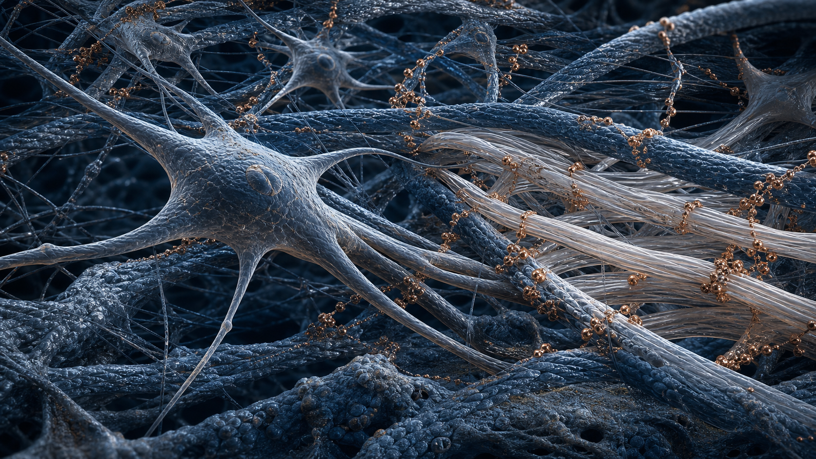
Scientific visualization of fibroblasts, collagen fibres, and copper-coloured molecular nodes

Copper Peptide Overview

Why GHK-Cu Is Everywhere in Skin and Tissue Science

Seven straightforward reasons GHK-Cu remains important in collagen, skin, hair, and tissue-remodeling research.

Compound overview • 4 minute read

Quick Take

GHK-Cu is a naturally occurring copper-binding tripeptide complex. Researchers study its effects on fibroblasts, collagen, extracellular matrix, blood-vessel growth, inflammation, oxidative stress, wound repair, and hair-follicle signaling.

Why It Gets Attention

GHK-Cu has a direct and visually understandable research story. Fibroblasts build collagen and extracellular matrix, while copper supports enzymes involved in tissue structure and repair.

Because the peptide intersects with several stages of remodeling, researchers can follow collagen expression, wound closure, vessel growth, inflammatory markers, antioxidant pathways, skin architecture, and follicle activity.

7 Key Areas Worth Knowing

The clearest themes are summarized below.

01

Collagen Production

GHK-Cu has been shown to stimulate collagen synthesis in cultured fibroblasts. Researchers examine collagen expression, procollagen markers, and organization of the extracellular matrix.

02

Wound Repair

Copper-peptide research is strongly connected with rebuilding damaged tissue. Wound closure, collagen deposition, re-epithelialization, and tissue strength are common endpoints.

03

Skin Structure and Elasticity

Collagen, elastin, and glycosaminoglycans help determine skin architecture. GHK-Cu models examine firmness, matrix quality, dermal organization, and age-related structural change.

04

Angiogenesis

New vessels support growing and repairing tissue. Researchers study endothelial-cell activity, vessel formation, and delivery of oxygen and nutrients to the repair site.

05

Inflammatory Balance

GHK-Cu has shown anti-inflammatory activity in several preclinical models. Cytokines, NF-kappaB signaling, inflammatory-cell activity, and tissue swelling can be measured.

06

Antioxidant Defense

Copper-peptide research also examines protection from oxidative stress. Reactive oxygen species, glutathione, antioxidant capacity, and Nrf2-related signaling are useful endpoints.

07

Hair-Follicle Signaling

GHK-Cu appears in hair-growth and follicle-viability research. Researchers examine follicle activity, growth-factor signaling, blood supply, and transition into active growth phases.

Why GHK-Cu Stands Out

The Collagen Signal Is Direct

A classic fibroblast study reported increased collagen synthesis with GHK-Cu. Later cell and tissue models have continued to examine type I, IV, and VII collagen, making matrix production a central theme.

Copper Adds Functional Biology

GHK binds copper and can help deliver it within biological systems. Copper is relevant to enzymes involved in connective-tissue crosslinking, antioxidant activity, and cellular metabolism.

Repair Includes More Than Collagen

A strong tissue-repair model also needs blood vessels and controlled inflammation. GHK-Cu research combines matrix production with angiogenesis, oxidative-stress control, and inflammatory signaling.

Skin and Hair Models Are Easy to Measure

Dermal thickness, collagen expression, wound closure, follicle stage, and vessel density are visible endpoints. That makes GHK-Cu especially engaging for aesthetic and regenerative research.

What Can Be Measured

These highlighted areas are most useful when treated as connected but separate endpoints. A controlled comparison can track collagen production, wound repair, skin structure and elasticity, angiogenesis, inflammatory balance, antioxidant defense, and hair-follicle signaling at planned time points. This turns a broad question into clear observations and shows which part of the compound’s profile changes most strongly.

A strong design also connects each outcome with the biology behind it. Measurements can be paired with markers related to the collagen signal is direct, copper adds functional biology, repair includes more than collagen, and skin and hair models are easy to measure. Combining visible or functional results with mechanism-focused data makes the positive findings easier to interpret, repeat, and compare across the available cell, animal, and clinical literature.

Evidence and Limitations

Much of the GHK-Cu evidence comes from cell, ex-vivo, and animal models, and delivery method can substantially affect results. Conclusions should remain tied to the tested formulation and model.

The Bottom Line

GHK-Cu stands out because its positive research areas connect directly with visible tissue structure. Collagen, wound repair, skin architecture, angiogenesis, inflammation, oxidative stress, and hair-follicle signaling create a coherent and highly engaging research profile.

Sources

  1. GHK-Cu stimulation of collagen synthesis in fibroblasts.
  2. Copper-peptide hydrogel and wound healing in mice.
  3. GHK-Cu, inflammation, and oxidative-stress signaling.

Related Resources

Review GHK-Cu 50mg or Open the Research Protocol

Research-use disclaimer: This article is for laboratory research education only. It does not provide medical advice or establish safety, efficacy, or suitability. Products discussed are not intended for human consumption, therapeutic use, diagnostic use, or veterinary use.

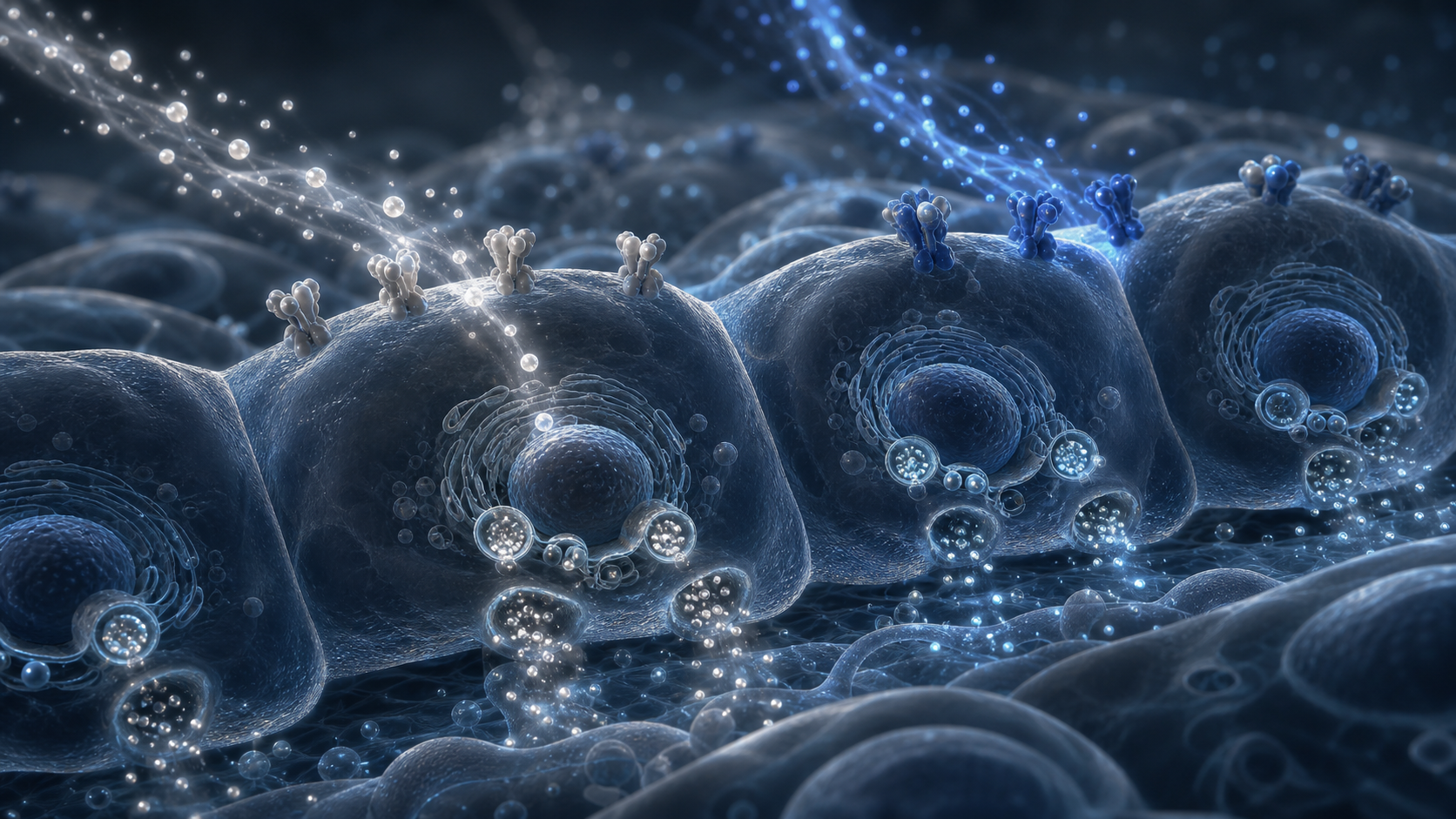
Posted on Leave a comment

Why CJC-1295 and Ipamorelin Are Often Paired

Two rowers smiling as they carry a red racing shell beside a bright lake

Peptide Overview

Why CJC-1295 and Ipamorelin Are Often Paired

Seven straightforward reasons this dual-pathway blend attracts attention in growth hormone, recovery, and metabolic research.

Compound overview • 4 minute read

Quick Take

CJC-1295 and Ipamorelin approach growth hormone signaling from two different directions. CJC-type compounds support GHRH-receptor signaling, while Ipamorelin activates the growth hormone secretagogue receptor. Together, they create a practical model for studying GH release and its wider effects.

Why the Combination Gets Attention

The attraction is simple: two compounds, two complementary signals. CJC-type compounds work on the GHRH side of the growth hormone axis. Ipamorelin works on the GHS-R1a side. Researchers can therefore examine how the combined signal compares with either pathway studied on its own.

That makes the blend relevant to GH pulse behavior, IGF-1 activity, protein turnover, tissue remodeling, energy use, and other downstream markers.

7 Key Areas Worth Knowing

Research interest in CJC-1295 + Ipamorelin generally centers on seven positive areas connected with GH and IGF-1 biology.

01

Muscle and Tissue Recovery

Growth hormone and IGF-1 support protein turnover and tissue remodeling. Researchers study whether coordinated GH-axis signaling can improve recovery markers after controlled physical stress.

02

Fat Metabolism

Growth hormone signaling is closely connected with lipolysis, the process of releasing stored fat for energy. This makes body-composition and energy-use markers a major area of interest.

03

Cellular Repair and Healthy-Aging Research

The GH and IGF-1 axis influences cellular maintenance, regeneration, and protein synthesis. Researchers examine these pathways when studying age-related changes in repair capacity and tissue quality.

04

Bone Strength and Remodeling

GH and IGF-1 participate in bone formation and mineral remodeling. Research models may track osteoblast activity, bone-turnover markers, and changes in mineral density.

05

Sleep-Linked GH Rhythms

Natural GH release is closely tied to deep sleep and overnight hormone pulses. Researchers study whether changes in pulse timing and amplitude correspond with sleep-quality and circadian markers.

06

Collagen and Connective-Tissue Support

GH and IGF-1 pathways are linked with fibroblast activity and collagen production. This supports research involving skin structure, tendons, ligaments, and broader connective-tissue remodeling.

07

Immune-System Signaling

The GH and IGF-1 axis also interacts with immune-cell development and signaling. Researchers examine these connections when studying immune balance, cellular communication, and recovery responses.

Why the Blend Stands Out

Two Complementary GH-Release Pathways

The blend does more than place two GH-related compounds in the same vial. CJC-type compounds support the GHRH-receptor pathway, which signals the pituitary to produce and release growth hormone. Ipamorelin activates GHS-R1a, a separate receptor involved in triggering GH release.

That dual-pathway design gives researchers a broader GH-axis model and makes direct comparisons with either compound alone especially useful.

Clear GH and IGF-1 Signaling

CJC-1295 has direct human research showing increased GH and IGF-1 activity. In controlled studies, the long-acting DAC form produced sustained, dose-dependent increases in both markers.

This gives researchers several straightforward measurements: mean GH concentration, IGF-1 response, duration of signal, and change from baseline. These markers help connect receptor activity with the recovery, tissue, and metabolic areas described above.

Preserved GH Pulse Dynamics

Growth hormone is naturally released in pulses rather than at one constant level. A human CJC-1295 study found that mean and trough GH increased while pulsatile secretion remained present.

Pulse timing, peak-to-trough behavior, and downstream IGF-1 response therefore remain important research endpoints. For shorter-acting CJC formats paired with Ipamorelin, pulse-oriented studies are an especially natural fit.

Ipamorelin’s Selective Profile

Ipamorelin was developed as a selective growth hormone secretagogue. In early animal research, it stimulated GH without the same significant ACTH and cortisol increases observed with GHRP-2 and GHRP-6 in the studied model.

That selective profile is one reason Ipamorelin remains popular in GH-axis research and is frequently paired with a CJC-type compound.

Evidence and Limitations

Published research often evaluates the two components separately, and long-acting CJC-1295 with DAC differs from shorter-acting no-DAC material. Results should be interpreted according to the compound identity and research model being studied.

The Bottom Line

CJC-1295 + Ipamorelin stands out because the combination is easy to understand and relevant to a wide range of positive research areas. Its dual-pathway design supports investigation into GH release, recovery, fat metabolism, cellular repair, bone remodeling, sleep-linked hormone rhythms, collagen production, and immune signaling.

Sources

  1. Teichman et al. CJC-1295 effects on GH and IGF-1 in healthy adults.
  2. Ionescu and Frohman. GH pulsatility during CJC-1295 stimulation.
  3. Raun et al. Selective GH-secretagogue activity of Ipamorelin.

Related Resources

Review the Blend or Open the Research Protocol

Research-use disclaimer: This article is for laboratory research education only. It does not provide medical advice or establish safety, efficacy, or suitability. Products discussed are not intended for human consumption, therapeutic use, diagnostic use, or veterinary use.

Posted on Leave a comment

Beyond Appetite: Seven Questions Around Semaglutide

Woman preparing a balanced lunch in a bright modern kitchen

Metabolic Peptide Overview

Beyond Appetite: Seven Questions Around Semaglutide

Seven straightforward reasons Semaglutide remains the reference point for GLP-1 receptor research.

Compound overview • 4 minute read

Quick Take

Semaglutide is a GLP-1 receptor agonist with extensive human research. Its focused one-receptor design makes it a useful reference model for appetite, energy intake, glucose regulation, body weight, cardiovascular outcomes, kidney markers, and liver-fat research.

Why It Gets Attention

Semaglutide provides a clean GLP-1 baseline. Researchers can compare it with dual agonists such as Tirzepatide and triple agonists such as Retatrutide to see what changes when additional receptors are added.

Large randomized trials have measured body weight, appetite, food intake, glucose, cardiovascular events, kidney outcomes, liver fat, and physical-function markers.

7 Key Areas Worth Knowing

The clearest themes are summarized below.

01

Body-Weight Reduction

The STEP 1 trial reported sustained, clinically meaningful body-weight reductions. Weight, waist circumference, body-mass index, and defined response targets are common research endpoints.

02

Reduced Hunger and Energy Intake

Controlled studies have reported lower hunger, greater fullness, fewer cravings, and reduced food intake. These measurements help explain the body-weight signal.

03

Glucose Regulation

GLP-1 signaling supports glucose-dependent insulin release and lower glucagon activity. Researchers follow HbA1c, fasting glucose, post-meal glucose, and insulin-response markers.

04

Cardiovascular Outcomes

The SELECT trial reported fewer major cardiovascular events in the studied population. This makes cardiovascular-risk markers and event outcomes an important research area.

05

Kidney-Outcome Research

SELECT and FLOW analyses reported positive kidney-related outcomes in defined clinical populations. Researchers examine eGFR, albuminuria, kidney events, and cardiovascular-kidney interactions.

06

Liver-Fat and Metabolic-Liver Research

Semaglutide studies have examined liver fat, liver enzymes, and steatohepatitis-related endpoints. This expands the research story beyond body weight and glucose.

07

Physical Function and Cardiometabolic Health

STEP 1 reported improvement in cardiometabolic risk factors and participant-reported physical functioning. Blood pressure, lipids, waist size, and functional measures add useful context.

Why Semaglutide Stands Out

A Focused GLP-1 Model

Semaglutide primarily represents GLP-1 receptor research. That focused mechanism makes cause-and-effect interpretation simpler than with multi-receptor compounds.

Appetite Data Explain the Weight Signal

Studies have directly measured energy intake and eating behavior. Lower hunger, greater satiety, fewer cravings, and reduced calorie intake provide a practical mechanism for observed body-weight changes.

Large Outcome Trials Add Depth

Semaglutide research includes more than short metabolic trials. SELECT and FLOW evaluate cardiovascular and kidney outcomes over longer periods and in large populations.

It Anchors Receptor Comparisons

Semaglutide is the logical baseline for comparing newer metabolic peptides. Researchers can add GIP or glucagon activity and examine which additional signals change.

What Can Be Measured

These highlighted areas are most useful when treated as connected but separate endpoints. A controlled comparison can track body-weight reduction, reduced hunger and energy intake, glucose regulation, cardiovascular outcomes, kidney-outcome research, liver-fat and metabolic-liver research, and physical function and cardiometabolic health at planned time points. This turns a broad question into clear observations and shows which part of the compound’s profile changes most strongly.

A strong design also connects each outcome with the biology behind it. Measurements can be paired with markers related to a focused glp-1 model, appetite data explain the weight signal, large outcome trials add depth, and it anchors receptor comparisons. Combining visible or functional results with mechanism-focused data makes the positive findings easier to interpret, repeat, and compare across the available cell, animal, and clinical literature.

Evidence and Limitations

Published clinical findings use regulated Semaglutide formulations and defined treatment protocols. They do not establish the performance or suitability of a separate research-use material.

The Bottom Line

Semaglutide remains a foundational metabolic-research peptide because the GLP-1 mechanism is focused and the human evidence is extensive. Weight, appetite, glucose, cardiovascular outcomes, kidney markers, liver fat, and physical function all fit within one well-defined research framework.

Sources

  1. STEP 1 once-weekly Semaglutide trial.
  2. SELECT cardiovascular outcomes trial.
  3. SELECT long-term kidney-outcomes analysis.

Related Resources

Review Semaglutide 20mg or Open the Research Protocol

Research-use disclaimer: This article is for laboratory research education only. It does not provide medical advice or establish safety, efficacy, or suitability. Products discussed are not intended for human consumption, therapeutic use, diagnostic use, or veterinary use.
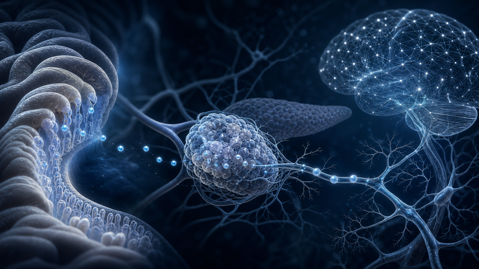
Posted on Leave a comment

Why NAD+ Sits at the Center of Cellular Energy

Scientific visualization of mitochondrial energy exchange and cellular redox cofactors

Cellular-Energy Overview

Why NAD+ Sits at the Center of Cellular Energy

Six straightforward reasons NAD+ remains central to cellular-energy, mitochondrial, and healthy-aging research.

Compound overview • 4 minute read

Quick Take

NAD+ is not a peptide; it is an essential cellular coenzyme. It transfers electrons during energy production and is consumed by enzymes involved in DNA repair, stress response, gene regulation, and cellular maintenance.

Why It Gets Attention

NAD+ sits near the center of cellular metabolism. Every cell needs it to move energy through glycolysis, the citric-acid cycle, and oxidative phosphorylation.

Researchers also track NAD+ because it supports PARP enzymes, sirtuins, redox balance, mitochondrial signaling, and responses to metabolic or age-related stress.

6 Key Areas Worth Knowing

The clearest themes are summarized below.

01

Cellular Energy Production

NAD+ carries electrons needed to convert nutrients into usable energy. ATP production, oxygen consumption, and metabolic flux are direct research endpoints.

02

Redox Balance

The NAD+/NADH ratio helps cells balance oxidation and reduction reactions. Researchers study how that ratio changes during exercise, fasting, oxidative stress, and metabolic dysfunction.

03

DNA-Repair Activity

PARP enzymes use NAD+ while responding to DNA damage. This connects NAD+ availability with genome maintenance, repair signaling, and cellular survival under stress.

04

Sirtuin Signaling

Sirtuins are NAD-dependent enzymes involved in metabolism and stress response. Researchers examine gene regulation, mitochondrial adaptation, inflammation, and cellular maintenance.

05

Muscle and Metabolic Research

Skeletal muscle has high energy demands and a large mitochondrial network. NAD+ studies track muscle metabolites, insulin sensitivity, fatigue, strength, and exercise efficiency.

06

Healthy-Aging Research

NAD+ metabolism changes with age and metabolic stress in several models. This supports research into resilience, repair capacity, mitochondrial health, and age-related functional decline.

Why NAD+ Stands Out

It Connects Nutrients to ATP

NAD+ accepts and transfers electrons as carbohydrates, fats, and amino acids are processed. That basic role makes it relevant to nearly every energy-demanding tissue.

It Is Both a Cofactor and a Substrate

NAD+ does more than carry electrons. PARPs, sirtuins, and other enzymes consume it while regulating DNA repair, stress responses, and gene activity.

Human Studies Can Measure the NAD Metabolome

Controlled precursor trials have shown that NAD-related metabolites can be raised in blood or muscle. These studies also show why higher NAD markers do not automatically guarantee the same functional outcome in every population.

It Anchors Mitochondrial Research

NAD+ availability, NADH oxidation, and ATP production can be studied together. That gives researchers a structured way to connect cellular chemistry with whole-tissue function.

What Can Be Measured

These highlighted areas are most useful when treated as connected but separate endpoints. A controlled comparison can track cellular energy production, redox balance, dna-repair activity, sirtuin signaling, muscle and metabolic research, and healthy-aging research at planned time points. This turns a broad question into clear observations and shows which part of the compound’s profile changes most strongly.

A strong design also connects each outcome with the biology behind it. Measurements can be paired with markers related to it connects nutrients to atp, it is both a cofactor and a substrate, human studies can measure the nad metabolome, and it anchors mitochondrial research. Combining visible or functional results with mechanism-focused data makes the positive findings easier to interpret, repeat, and compare across the available cell, animal, and clinical literature.

Evidence and Limitations

Many human studies raise NAD+ through precursors such as NR, NMN, or niacin rather than evaluating a direct NAD+ research vial. Findings should not be transferred between formats without confirming exposure and metabolism.

The Bottom Line

NAD+ stands out because it links energy production with repair and stress-response systems. Cellular energy, redox balance, mitochondria, DNA repair, sirtuins, muscle metabolism, and healthy aging all connect through one essential coenzyme.

Sources

  1. Nicotinamide riboside and the aged human muscle NAD metabolome.
  2. NMN supplementation and blood NAD+ in older men.
  3. Niacin, NAD+ deficiency, and mitochondrial myopathy.

Related Resources

Review NAD+ 500mg or Open the Research Protocol

Research-use disclaimer: This article is for laboratory research education only. It does not provide medical advice or establish safety, efficacy, or suitability. Products discussed are not intended for human consumption, therapeutic use, diagnostic use, or veterinary use.
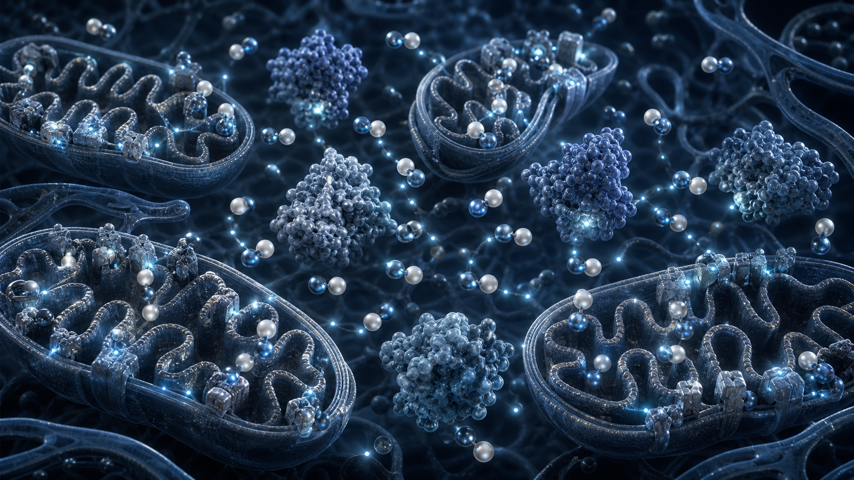
Posted on Leave a comment

Tesamorelin Beyond the Scale: Visceral Fat, Liver Fat, and More

Smiling mature boxer resting between rounds in a bright training studio

Growth-Hormone Axis Overview

Tesamorelin Beyond the Scale: Visceral Fat, Liver Fat, and More

Six clear reasons Tesamorelin remains a major compound in visceral-fat, liver-fat, body-composition, and growth-hormone-axis research.

Compound overview • 4 minute read

Quick Take

Tesamorelin is a growth hormone-releasing hormone analog with substantial human clinical data in specific populations. Researchers study its effects on pulsatile GH release, IGF-1, visceral adipose tissue, liver fat, waist measurements, lipids, and inflammatory markers.

Why It Gets Attention

Tesamorelin has a direct and highly measurable research profile. Imaging studies can separate visceral fat from subcutaneous fat, while blood measurements can track IGF-1, triglycerides, glucose regulation, liver markers, and inflammation.

Unlike broader weight-focused models, Tesamorelin research often asks where body-composition change occurs. That makes CT, MRI, magnetic-resonance spectroscopy, waist measurements, and tissue-specific fat analysis especially important.

6 Key Areas Worth Knowing

The clearest themes are summarized below.

01

Visceral-Fat Reduction

Randomized trials have reported meaningful reductions in visceral adipose tissue in studied populations. CT and MRI measurements provide direct, location-specific outcomes rather than relying on scale weight alone.

02

Liver-Fat Research

Tesamorelin studies have reported reductions in hepatic fat measured by imaging. Liver-fat fraction, liver enzymes, fibrosis-related markers, and histological features can be followed.

03

Selective Body-Composition Change

Tesamorelin research is notable for distinguishing visceral fat from subcutaneous and limb fat. That selectivity helps researchers test whether changes are regional rather than simply reflecting overall weight loss.

04

Lipid Metabolism

Clinical studies have followed triglycerides and other lipid markers alongside body-composition change. Triglycerides, cholesterol fractions, liver lipids, and fatty-acid metabolism add depth to imaging results.

05

Inflammatory and Cardiometabolic Markers

Some studies have reported improvements in C-reactive protein and vascular measurements. Inflammatory markers, carotid measurements, blood pressure, and glucose endpoints can be tracked together.

06

GH and IGF-1 Signaling

Tesamorelin works through the natural growth-hormone-releasing hormone receptor pathway. Pulsatile GH release, IGF-1, downstream metabolic markers, and pituitary responsiveness are central mechanistic endpoints.

Why Tesamorelin Stands Out

Imaging Shows Where Change Occurs

The strongest Tesamorelin studies use CT, MRI, or spectroscopy instead of relying only on body weight. Researchers can separate visceral, subcutaneous, abdominal, and liver fat to build a much clearer metabolic picture.

The GH Axis Remains Physiologically Organized

As a GHRH analog, Tesamorelin stimulates the pituitary side of the GH axis. That mechanism differs from providing growth hormone directly and allows researchers to study pulsatile release and IGF-1 response.

Visceral and Liver Fat Can Be Studied Together

Randomized research has measured both abdominal visceral adipose tissue and hepatic fat. This creates a useful model for examining how regional fat stores relate to broader metabolic markers.

Human Data Provide Clear Benchmarks

Tesamorelin has more controlled human evidence than most research peptides. The available trials provide defined imaging, body-composition, lipid, and endocrine endpoints for comparison.

What Can Be Measured

These highlighted areas are most useful when treated as connected but separate endpoints. A controlled comparison can track visceral-fat reduction, liver-fat research, selective body-composition change, lipid metabolism, inflammatory and cardiometabolic markers, and gh and igf-1 signaling at planned time points. This turns a broad question into clear observations and shows which part of the compound’s profile changes most strongly.

A strong design also connects each outcome with the biology behind it. Measurements can be paired with markers related to imaging shows where change occurs, the gh axis remains physiologically organized, visceral and liver fat can be studied together, and human data provide clear benchmarks. Combining visible or functional results with mechanism-focused data makes the positive findings easier to interpret, repeat, and compare across the available cell, animal, and clinical literature.

Evidence and Limitations

Tesamorelin is the active molecule in a regulated prescription product for a specific indication. Results from that formulation and population do not automatically validate a separate research-use vial.

The Bottom Line

Tesamorelin stands out because its positive research areas are visible and measurable. Visceral fat, liver fat, waist measurements, regional body composition, lipids, inflammation, GH release, and IGF-1 can all be studied within one endocrine framework.

Sources

  1. Tesamorelin, visceral fat, and body-composition outcomes.
  2. Randomized Tesamorelin trial measuring visceral and liver fat.
  3. Tesamorelin and liver-fat research in a randomized trial.

Related Resources

Review Tesamorelin 15mg or Open the Research Protocol

Research-use disclaimer: This article is for laboratory research education only. It does not provide medical advice or establish safety, efficacy, or suitability. Products discussed are not intended for human consumption, therapeutic use, diagnostic use, or veterinary use.

Posted on Leave a comment

Inside KLOW Blend: Four Angles in One Formula

Colorful scientific visualization of collagen, migrating cells, signalling particles, and epithelial tissue

Multi-Peptide Repair Overview

Inside KLOW Blend: Four Angles in One Formula

Six clear reasons the BPC-157, TB-500, KPV, and GHK-Cu combination creates a broad repair and tissue-remodeling research model.

Compound overview • 4 minute read

Quick Take

KLOW combines 10mg BPC-157, 10mg TB-500, 10mg KPV, and 50mg GHK-Cu in one research vial. The four components bring together collagen, cell migration, inflammatory signaling, gastrointestinal integrity, angiogenesis, and tissue-remodeling questions.

Why It Gets Attention

KLOW is interesting because repair is not a single biological event. Cells must move into the area, inflammatory signaling must remain organized, blood vessels must support the tissue, and collagen must be produced and remodeled into a functional structure.

The blend lets researchers examine those stages together while using single-component control groups to determine whether BPC-157, thymosin beta-4-related material, KPV, or GHK-Cu is driving a particular result.

6 Key Areas Worth Knowing

The clearest themes are summarized below.

01

Collagen and Matrix Remodeling

GHK-Cu and BPC-157 research both connect with collagen biology. Researchers can follow fibroblast activity, collagen expression, extracellular-matrix organization, crosslinking, and mechanical strength.

02

Cell Migration

Thymosin beta-4-related research emphasizes actin regulation and movement of repair-associated cells. Migration assays, epithelial coverage, myoblast recruitment, and endothelial movement help show how rebuilding begins.

03

Inflammatory Balance

KPV gives the blend a recognizable anti-inflammatory research component. NF-kappaB, MAP kinase signaling, cytokine release, immune-cell activity, and tissue swelling can all be measured.

04

Gut and Barrier Integrity

Both KPV and BPC-157 appear frequently in gastrointestinal research models. Barrier resistance, tight-junction proteins, epithelial wound closure, mucosal structure, and inflammatory markers are practical endpoints.

05

Angiogenesis and Circulation

Repairing tissue needs oxygen, nutrients, and a stable local blood supply. The blend supports research involving endothelial activity, new-vessel formation, vascular density, and blood-flow-related signaling.

06

Skin and Connective-Tissue Research

The high GHK-Cu component makes visible tissue remodeling especially relevant. Researchers can examine dermal structure, collagen density, wound appearance, elasticity markers, and connective-tissue organization.

Why the KLOW Blend Stands Out

Four Components Cover Different Repair Stages

BPC-157 emphasizes local tissue response, TB-500 emphasizes cell movement, KPV emphasizes inflammatory pathways, and GHK-Cu emphasizes matrix remodeling. That division gives the blend a clear rationale without pretending every component does the same job.

KPV Adds a Distinct Barrier-Research Angle

KPV studies have reported reduced inflammatory signaling and improved epithelial barrier measurements. This separates KLOW from simpler recovery blends and makes intestinal or skin-barrier models particularly relevant.

GHK-Cu Makes Collagen Easy to Track

GHK-Cu is the largest component by mass and has a long research history involving collagen and extracellular matrix. Fibroblast activity and structural tissue measurements provide concrete outcomes alongside molecular markers.

Controls Can Separate Combination Effects

A well-designed blend study can compare KLOW with each individual component and with simpler combinations. That structure helps show whether the full blend produces additive, overlapping, or component-specific changes.

What Can Be Measured

These highlighted areas are most useful when treated as connected but separate endpoints. A controlled comparison can track collagen and matrix remodeling, cell migration, inflammatory balance, gut and barrier integrity, angiogenesis and circulation, and skin and connective-tissue research at planned time points. This turns a broad question into clear observations and shows which part of the compound’s profile changes most strongly.

A strong design also connects each outcome with the biology behind it. Measurements can be paired with markers related to four components cover different repair stages, kpv adds a distinct barrier-research angle, ghk-cu makes collagen easy to track, and controls can separate combination effects. Combining visible or functional results with mechanism-focused data makes the positive findings easier to interpret, repeat, and compare across the available cell, animal, and clinical literature.

Evidence and Limitations

The exact four-component KLOW Blend has not been evaluated in controlled human trials. Research on individual ingredients supports study questions but cannot establish the combined material’s safety, interactions, or performance.

The Bottom Line

KLOW stands out because its four components create a full repair sequence rather than a one-pathway story. Cell migration, inflammatory balance, barrier integrity, angiogenesis, collagen production, and tissue remodeling can all be followed in the same structured model.

Sources

  1. BPC-157 and tendon-healing research.
  2. KPV uptake and intestinal inflammatory signaling.
  3. Copper-peptide tissue-remodeling research.

Related Resources

Review KLOW Blend or Open the Research Protocol

Research-use disclaimer: This article is for laboratory research education only. It does not provide medical advice or establish safety, efficacy, or suitability. Products discussed are not intended for human consumption, therapeutic use, diagnostic use, or veterinary use.

Posted on Leave a comment

Epitalon and the Longevity Question

Silver-haired woman riding a dark horse beside a cool coastal field

Longevity Peptide Overview

Epitalon and the Longevity Question

Five straightforward reasons Epitalon remains a recognizable compound in telomere, pineal, cellular-aging, and longevity research.

Compound overview • 4 minute read

Quick Take

Epitalon is a synthetic tetrapeptide with the sequence Ala-Glu-Asp-Gly. Its research profile centers on telomerase activity, telomere length, antioxidant defenses, pineal signaling, circadian biology, and age-related cellular change.

Why It Gets Attention

Epitalon has a clear research identity because it connects a very small peptide with large questions about cellular aging. Early cell studies reported telomerase activation and telomere elongation, while later work expanded the discussion to antioxidant enzymes and neuroendocrine signaling.

Researchers can follow direct molecular endpoints such as hTERT expression, telomerase activity, telomere length, reactive oxygen species, antioxidant enzymes, melatonin-related signaling, cellular senescence, and survival in aging models.

5 Key Areas Worth Knowing

The clearest themes are summarized below.

01

Telomerase and Telomere-Length Research

Epitalon research is best known for reported telomerase activation and telomere elongation in cultured human cells. Researchers can measure hTERT expression, enzyme activity, telomere length, replication capacity, and the timing of the cellular response.

02

Antioxidant Defense

Pineal-peptide research has linked Epitalon with stronger antioxidant activity in older animal models. Superoxide dismutase, glutathione-related enzymes, reactive oxygen species, and lipid oxidation are useful measurements.

03

Circadian and Melatonin Signaling

Epitalon’s pineal origin keeps melatonin and biological timing central to its research profile. Studies examine light-cycle disruption, melatonin production, daily rhythmicity, and age-related changes in circadian signaling.

04

Neuroendocrine Regulation

The peptide is studied as a signal that may connect the pineal gland with broader hormonal regulation. Researchers follow hypothalamic signaling, reproductive timing, stress-related pathways, and age-dependent neuroendocrine changes.

05

Longevity Models

Animal research has made lifespan and health-span questions a major part of Epitalon’s reputation. Survival curves, age-related function, tumor incidence, physical endurance, and late-life resilience have all appeared as endpoints.

Why Epitalon Stands Out

The Telomerase Signal Is Direct

A foundational human-fibroblast study reported expression of the catalytic telomerase subunit and telomere elongation. That gives Epitalon a specific molecular mechanism that can be tested with established laboratory methods rather than relying only on broad aging observations.

Newer Cell Work Expands the Comparison

Recent research has examined normal epithelial cells, fibroblasts, and cancer cell lines side by side. This allows researchers to compare hTERT-driven telomerase activity with alternative telomere-lengthening pathways across different cell types.

Antioxidant Biology Adds Another Layer

Older-rat studies reported changes in antioxidant and antiradical activity. Following enzymes such as superoxide dismutase and glutathione peroxidase helps connect cellular-aging questions with protection from oxidative stress.

Pineal Biology Keeps the Model Broad

Melatonin and circadian signaling give Epitalon a second recognizable research theme. This makes it possible to study molecular aging, biological timing, and neuroendocrine organization within one coherent program.

What Can Be Measured

These highlighted areas are most useful when treated as connected but separate endpoints. A controlled comparison can track telomerase and telomere-length research, antioxidant defense, circadian and melatonin signaling, neuroendocrine regulation, and longevity models at planned time points. This turns a broad question into clear observations and shows which part of the compound’s profile changes most strongly.

A strong design also connects each outcome with the biology behind it. Measurements can be paired with markers related to the telomerase signal is direct, newer cell work expands the comparison, antioxidant biology adds another layer, and pineal biology keeps the model broad. Combining visible or functional results with mechanism-focused data makes the positive findings easier to interpret, repeat, and compare across the available cell, animal, and clinical literature.

Evidence and Limitations

Epitalon evidence is dominated by cell and animal research, much of it from a limited group of investigators. Telomerase findings do not establish human longevity effects or the performance of a separate research vial.

The Bottom Line

Epitalon stands out because its research profile begins with one of aging biology’s most recognizable structures: the telomere. Telomerase, cellular replication, antioxidant defense, circadian signaling, neuroendocrine regulation, and longevity models create an unusually focused research story.

Sources

  1. Epitalon, telomerase activity, and telomere elongation in human cells.
  2. Antioxidant properties of Epitalon in older animal models.
  3. Epitalon and telomere-length pathways in human cell lines.

Related Resources

Review Epitalon 50mg or Open the Research Protocol

Research-use disclaimer: This article is for laboratory research education only. It does not provide medical advice or establish safety, efficacy, or suitability. Products discussed are not intended for human consumption, therapeutic use, diagnostic use, or veterinary use.
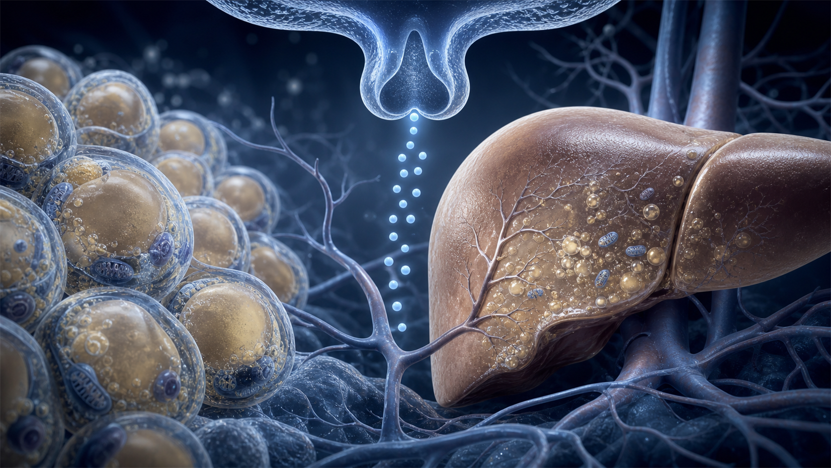
Posted on Leave a comment

TB-500 and Tissue Repair: Seven Signals Worth Watching

Gymnast holding a controlled strength position on still rings

Recovery Peptide Overview

TB-500 and Tissue Repair: Seven Signals Worth Watching

Seven clear reasons thymosin beta-4-related research remains important in cell migration, tissue remodeling, and recovery models.

Compound overview • 4 minute read

Quick Take

TB-500 is commonly discussed through thymosin beta-4 biology. That pathway is strongly connected with actin regulation, cell movement, blood-vessel development, inflammatory balance, and tissue-repair signaling.

Why It Gets Attention

The central idea is movement: repairing cells need to reach damaged tissue before rebuilding can begin. Thymosin beta-4 research focuses heavily on actin dynamics and the migration of epithelial cells, myoblasts, endothelial cells, and other repair-associated cells.

Researchers can therefore follow wound closure, new-vessel growth, tissue organization, inflammatory markers, apoptosis, and restoration of function across several models.

7 Key Areas Worth Knowing

The clearest themes are summarized below.

01

Cell Migration

Thymosin beta-4-related signaling helps organize actin, a structural protein cells use for movement. This makes cell migration one of the clearest and most practical TB-500 research themes.

02

Wound Closure

Faster movement of repair-associated cells can support re-epithelialization and tissue coverage. Researchers examine closure rate, epithelial organization, scarring, and restoration of normal structure.

03

Muscle-Recovery Signaling

Thymosin beta-4 expression rises after muscle injury in animal models. Studies have connected it with myoblast attraction, local cell communication, and early stages of muscle regeneration.

04

Angiogenesis

New vessels improve oxygen and nutrient delivery to rebuilding tissue. Thymosin beta-4 research has examined endothelial migration, vascular growth, and stabilization of developing vessel networks.

05

Inflammatory Balance

Controlled inflammation is necessary for repair, but excessive inflammation can slow it. Researchers study cytokines, inflammatory-cell movement, oxidative stress, and resolution of the repair response.

06

Cardiac-Tissue Repair

Preclinical cardiac research has linked thymosin beta-4 with cell survival and new-vessel development. These models examine myocardial protection, progenitor-cell activity, and vascular regeneration after injury.

07

Corneal and Epithelial Healing

Corneal models provide some of the clearest evidence for epithelial migration and wound closure. Research has followed re-epithelialization, reduced inflammatory disruption, and improved surface repair.

Why TB-500 Stands Out

Actin Dynamics Drive the Research Story

Actin gives cells shape and allows them to move. Thymosin beta-4 binds actin and influences how it is organized. That mechanism provides a straightforward explanation for the compound’s broad relevance to migration and repair.

Repair Cells Are Drawn Toward Injury

A mouse muscle study found increased thymosin beta-4 expression after injury and chemotactic effects on myoblasts. That makes recruitment of repair-associated cells an especially useful endpoint.

Vascular Growth Supports Rebuilding

Tissue cannot rebuild efficiently without circulation. Cardiac and endothelial research has linked thymosin beta-4 with neovascularization, vessel stabilization, and improved support for injured tissue.

Multiple Tissue Models Use the Same Core Mechanism

Muscle, cornea, heart, and skin models all rely on cell movement and structural remodeling. That shared biology is why thymosin beta-4-related research covers so many different tissues without losing a clear central theme.

What Can Be Measured

These highlighted areas are most useful when treated as connected but separate endpoints. A controlled comparison can track cell migration, wound closure, muscle-recovery signaling, angiogenesis, inflammatory balance, cardiac-tissue repair, and corneal and epithelial healing at planned time points. This turns a broad question into clear observations and shows which part of the compound’s profile changes most strongly.

A strong design also connects each outcome with the biology behind it. Measurements can be paired with markers related to actin dynamics drive the research story, repair cells are drawn toward injury, vascular growth supports rebuilding, and multiple tissue models use the same core mechanism. Combining visible or functional results with mechanism-focused data makes the positive findings easier to interpret, repeat, and compare across the available cell, animal, and clinical literature.

Evidence and Limitations

Published studies commonly evaluate full-length thymosin beta-4, while retail TB-500 naming may refer to related synthetic material. Compound identity should be confirmed before applying published findings.

The Bottom Line

TB-500 stands out because its research story is direct: cell movement supports repair. Actin regulation, wound closure, muscle signaling, angiogenesis, inflammatory balance, cardiac repair, and epithelial healing all connect back to that central mechanism.

Sources

  1. Thymosin beta-4 and myoblast migration after muscle injury.
  2. Thymosin beta-4 and cardiac neovascularization.
  3. Thymosin beta-4 and epithelial-cell migration.

Related Resources

Review TB-500 10mg or Open the Research Protocol

Research-use disclaimer: This article is for laboratory research education only. It does not provide medical advice or establish safety, efficacy, or suitability. Products discussed are not intended for human consumption, therapeutic use, diagnostic use, or veterinary use.