
Posted on Leave a comment

Ipamorelin Explained: The Selective GH-Release Story

Smiling swimmer relaxing at the edge of a sunlit turquoise lap pool

Growth-Hormone Secretagogue Overview

Ipamorelin Explained: The Selective GH-Release Story

Five straightforward reasons Ipamorelin remains important in selective growth-hormone release, ghrelin-receptor, and gastrointestinal-motility research.

Compound overview • 4 minute read

Quick Take

Ipamorelin is a synthetic pentapeptide and ghrelin-receptor agonist developed as a selective growth-hormone secretagogue. Its research profile combines GH release, pituitary selectivity, IGF-1 signaling, gastric motility, and comparison with other GH-axis compounds.

Why It Gets Attention

Ipamorelin became recognizable because early pharmacology separated growth-hormone release from broader pituitary-hormone effects. In animal models it stimulated GH while producing much smaller ACTH and cortisol responses than older secretagogues.

Its ghrelin-receptor activity also creates a second research path involving gastric smooth muscle, emptying, intestinal transit, and postoperative motility. That combination makes the compound broader than a simple GH-release model.
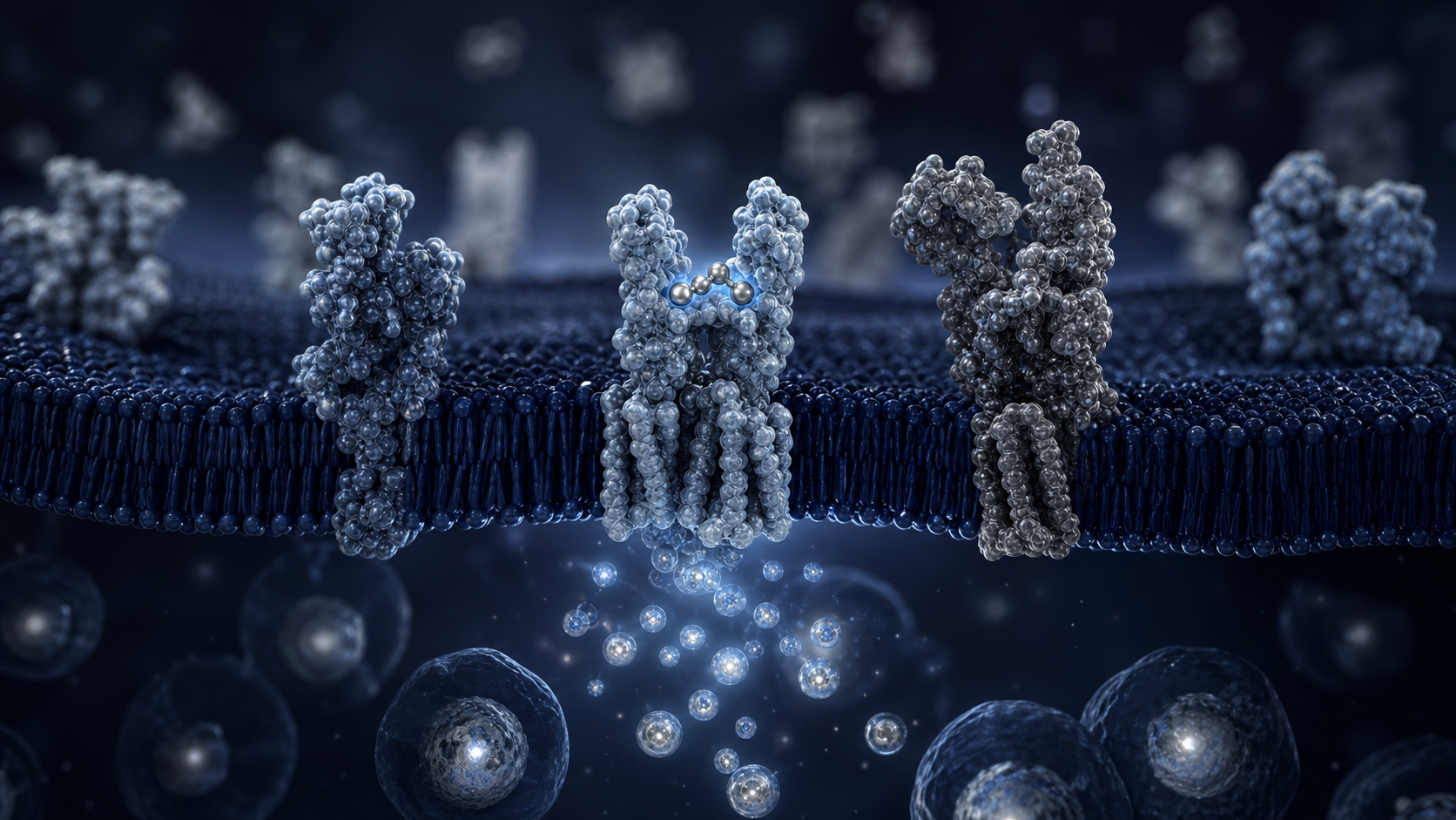
5 Key Areas Worth Knowing

The clearest themes are summarized below.

01

Selective Growth-Hormone Release

Ipamorelin was identified for potent GH release with a comparatively selective hormone profile in animal studies. GH pulse size, timing, pituitary response, ACTH, cortisol, and comparison with GHRH or older GHRPs are useful endpoints.

02

Ghrelin-Receptor Signaling

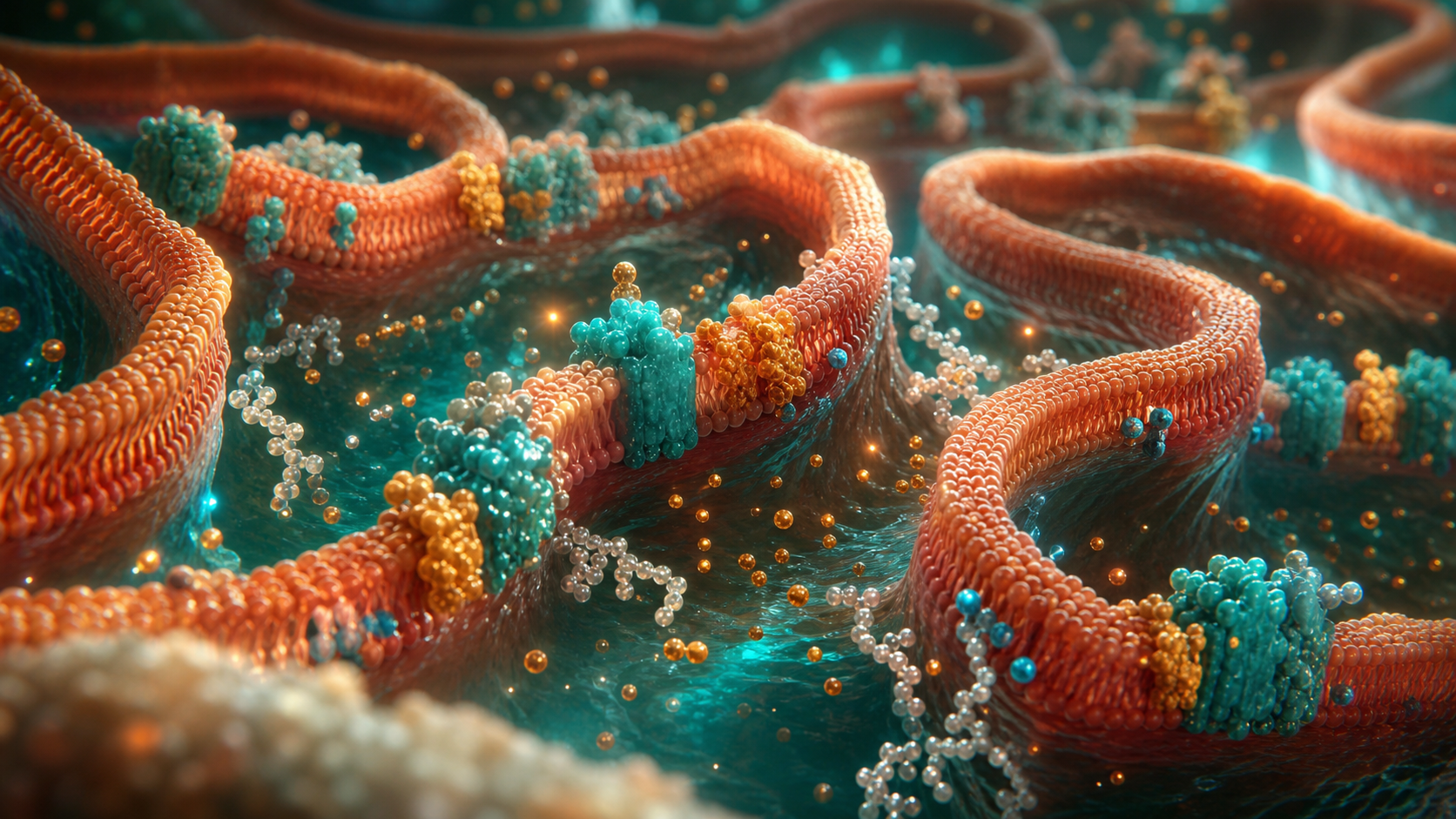
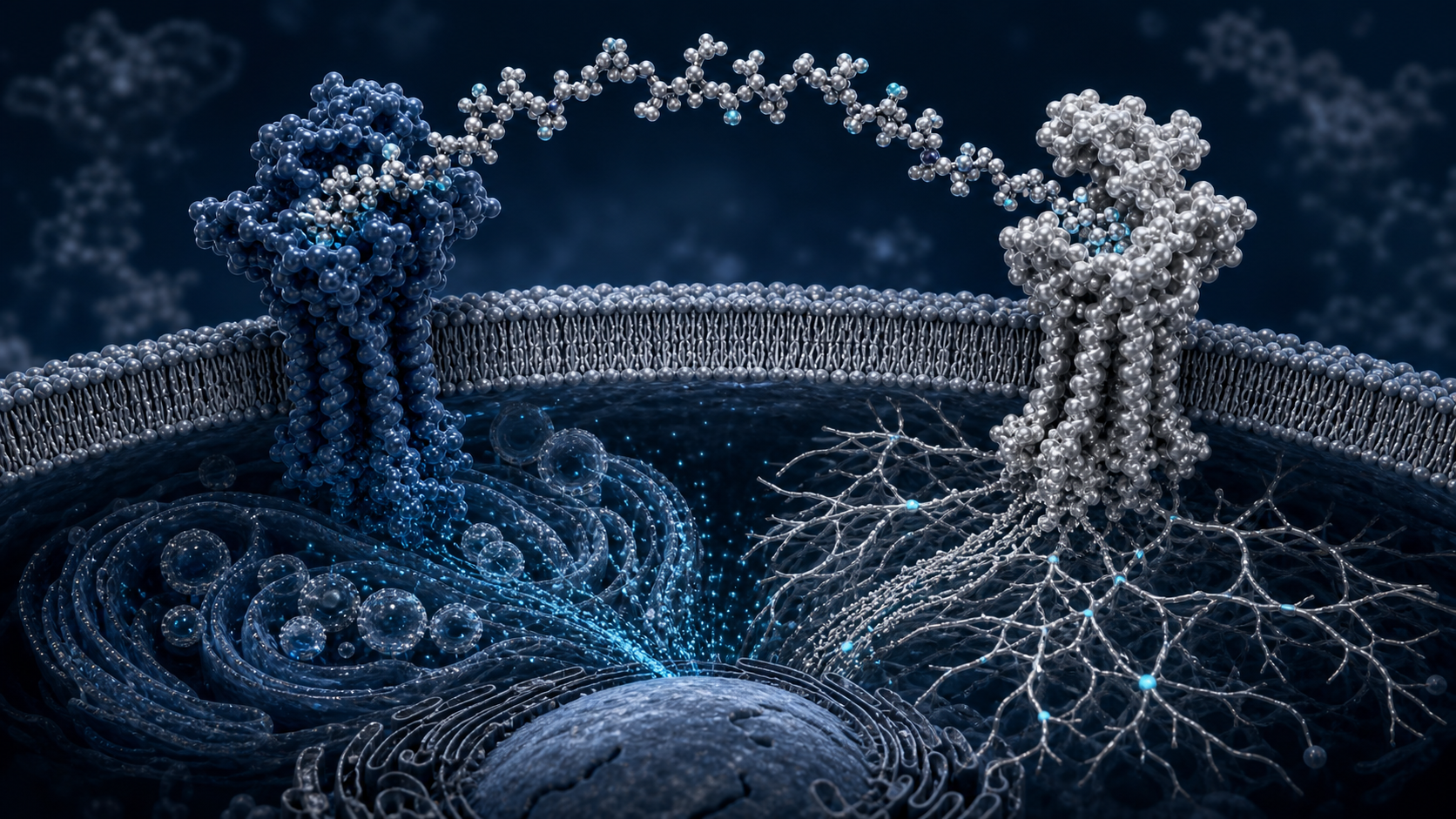
Ipamorelin activates the growth-hormone secretagogue receptor used by ghrelin. Receptor binding, downstream calcium signaling, pituitary-cell response, and tissue-specific activity can be measured.

03

IGF-1 and Anabolic Signaling

Growth-hormone release connects Ipamorelin with downstream IGF-1 research. Researchers can follow IGF-1, protein-synthesis pathways, collagen turnover, nitrogen balance, and tissue-growth markers.

04

Gastric Emptying

Rodent studies reported improved gastric emptying after surgery-induced dysmotility. Stomach retention, emptying rate, smooth-muscle contraction, and cholinergic signaling provide direct measurements.

05

Intestinal Transit Research

Ipamorelin has also been studied in postoperative intestinal-motility models. Transit time, bowel movement timing, fecal output, food intake, and restoration of gastrointestinal function can be followed.

Why Ipamorelin Stands Out

Selectivity Defines the Compound

The original pharmacology paper described Ipamorelin as the first selective growth-hormone secretagogue. Comparing GH with ACTH, cortisol, prolactin, thyroid, and gonadotropin responses gives researchers a clear way to test that profile.

The Ghrelin Receptor Explains Two Research Paths

Ghrelin-receptor activation affects both the pituitary and the gastrointestinal system. Ipamorelin can therefore be studied through hormone release and motility without treating those outcomes as unrelated.

Smooth-Muscle Measurements Are Direct

Postoperative models have measured stomach retention and isolated gastric-muscle contraction. These functional endpoints complement hormone measurements and show receptor activity at the tissue level.

It Supports Clear Head-to-Head Comparisons

Ipamorelin can be compared with GHRH analogs, GHRP-6, GHRP-2, ghrelin, and combination models. Those comparisons help separate potency, efficacy, receptor selectivity, and downstream hormone patterns.

What Can Be Measured

These highlighted areas are most useful when treated as connected but separate endpoints. A controlled comparison can track selective growth-hormone release, ghrelin-receptor signaling, igf-1 and anabolic signaling, gastric emptying, and intestinal transit research at planned time points. This turns a broad question into clear observations and shows which part of the compound’s profile changes most strongly.

A strong design also connects each outcome with the biology behind it. Measurements can be paired with markers related to selectivity defines the compound, the ghrelin receptor explains two research paths, smooth-muscle measurements are direct, and it supports clear head-to-head comparisons. Combining visible or functional results with mechanism-focused data makes the positive findings easier to interpret, repeat, and compare across the available cell, animal, and clinical literature.

Evidence and Limitations

Most Ipamorelin evidence is preclinical, and a human postoperative trial did not meet its primary efficacy endpoint. Research conclusions should remain tied to the tested material, model, and measured outcome.

The Bottom Line

Ipamorelin stands out because selectivity gives its research story a clear center. GH release, reduced pituitary spillover, ghrelin-receptor signaling, IGF-1, tissue-recovery questions, gastric emptying, and intestinal transit all fit within one receptor model.

Sources

  1. Ipamorelin as a selective growth-hormone secretagogue.
  2. Ipamorelin and intestinal transit in a postoperative model.
  3. Ipamorelin and gastric emptying in a rodent model.

Related Resources

Review Ipamorelin 10mg or Open the Research Protocol

Research-use disclaimer: This article is for laboratory research education only. It does not provide medical advice or establish safety, efficacy, or suitability. Products discussed are not intended for human consumption, therapeutic use, diagnostic use, or veterinary use.

Posted on Leave a comment

Two Recovery Signals, One Strategy: BPC-157 + TB-500

Two trail runners laughing while pausing to retie a shoe on a bright mountain trail

Recovery Blend Overview

Two Recovery Signals, One Strategy: BPC-157 + TB-500

Six straightforward reasons researchers pair BPC-157 tissue-response research with thymosin beta-4-related cell-migration research.

Compound overview • 4 minute read

Quick Take

BPC-157 + TB-500 combines two distinct recovery-research themes. BPC-157 is commonly studied around local tissue response, collagen, vascular signaling, and gastrointestinal protection, while thymosin beta-4-related research emphasizes actin, cell migration, and structural remodeling.

Why It Gets Attention

The blend is appealing because tissue repair requires more than one event. Cells must migrate, blood vessels must develop, collagen must organize, inflammation must settle, and the repaired structure must regain strength.

Pairing these compounds gives researchers one model for following several stages of recovery while still allowing comparison with single-compound control groups.

6 Key Areas Worth Knowing

The clearest themes are summarized below.

01

Tendon and Ligament Remodeling

BPC-157 research emphasizes tendon strength and collagen organization, while thymosin beta-4 biology emphasizes cell movement. Together, those themes support structured tendon and ligament research.

02

Cell Migration to Damaged Tissue

Repair cells must reach the target area before rebuilding can begin. Thymosin beta-4-related actin signaling makes migration a central endpoint in blend research.

03

Angiogenesis and Circulation

Both research categories intersect with vascular response. New-vessel formation can be studied alongside oxygen delivery, endothelial activity, and restoration of local circulation.

04

Inflammatory Balance

Recovery depends on a controlled inflammatory response. Blend research can track edema, inflammatory-cell activity, cytokine signaling, and transition into the rebuilding phase.

05

Collagen and Wound Strength

BPC-157 studies have reported collagen development and improved mechanical properties in animal models. The blend adds a complementary cell-migration framework to those structural endpoints.

06

Muscle and Soft-Tissue Recovery

Myoblast movement, vascular growth, collagen organization, and local tissue signaling all matter after physical stress. The combination brings those positive research themes into one model.

Why the Blend Stands Out

Two Different Repair Angles

BPC-157 and thymosin beta-4-related compounds are not duplicates. One is commonly studied for tissue protection, vascular signaling, and structural healing; the other is strongly tied to actin regulation and movement of repair-associated cells.

Repair Can Be Followed in Stages

A strong blend study can separate migration, inflammation, vascularization, collagen formation, and mechanical recovery. That staged approach helps researchers understand when the combination changes the repair process.

Single-Compound Controls Improve the Design

The blend becomes more informative when compared with BPC-157 alone and TB-500 alone. Those control groups help show whether an observed signal comes from one component or from the combination.

The Benefits Are Easy to Understand

The blend has a clear layman-level explanation. BPC-157 supports tissue-response research, while thymosin beta-4-related signaling supports movement and remodeling. Those functions naturally complement each other.

What Can Be Measured

These highlighted areas are most useful when treated as connected but separate endpoints. A controlled comparison can track tendon and ligament remodeling, cell migration to damaged tissue, angiogenesis and circulation, inflammatory balance, collagen and wound strength, and muscle and soft-tissue recovery at planned time points. This turns a broad question into clear observations and shows which part of the compound’s profile changes most strongly.

A strong design also connects each outcome with the biology behind it. Measurements can be paired with markers related to two different repair angles, repair can be followed in stages, single-compound controls improve the design, and the benefits are easy to understand. Combining visible or functional results with mechanism-focused data makes the positive findings easier to interpret, repeat, and compare across the available cell, animal, and clinical literature.

Evidence and Limitations

Published research generally evaluates BPC-157 and thymosin beta-4 separately. Direct evidence for the exact combined retail vial is limited, so component identity and comparison groups remain important.

The Bottom Line

BPC-157 + TB-500 stands out as a broad recovery-research blend. Tendon remodeling, cell migration, angiogenesis, inflammation, collagen strength, wound closure, and soft-tissue recovery provide a practical set of positive endpoints for structured laboratory study.

Sources

  1. BPC-157 and Achilles-tendon healing in rats.
  2. Thymosin beta-4 and myoblast migration.
  3. Thymosin beta-4 and vascular regeneration.

Related Resources

Review BPC-157 + TB-500 10mg+10mg or Open the Research Protocol

Research-use disclaimer: This article is for laboratory research education only. It does not provide medical advice or establish safety, efficacy, or suitability. Products discussed are not intended for human consumption, therapeutic use, diagnostic use, or veterinary use.
Posted on Leave a comment

Retatrutide in the Spotlight: What Triple-Agonist Studies Measure

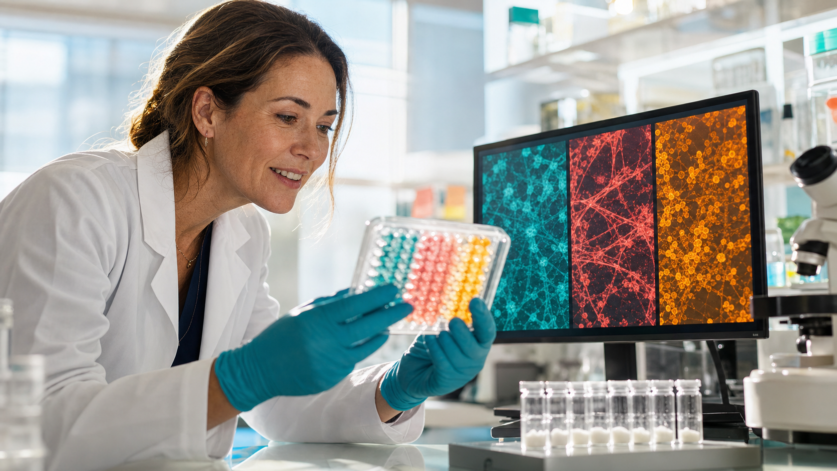
Scientist reviewing three colorful metabolic assay channels beside research vials

Metabolic Peptide Overview

Retatrutide in the Spotlight: What Triple-Agonist Studies Measure

Seven straightforward reasons the GLP-1, GIP, and glucagon triple-agonist model has become a major metabolic research topic.

Compound overview • 4 minute read

Quick Take

Retatrutide is a single peptide designed to activate three metabolic receptors: GLP-1, GIP, and glucagon. That triple-receptor profile allows researchers to examine appetite, glucose regulation, insulin sensitivity, energy use, body weight, and liver-fat biology together.

Why It Gets Attention

Most incretin research starts with one or two receptors. Retatrutide adds glucagon-receptor activity to the GLP-1 and GIP framework, creating a broader model for studying both energy intake and energy expenditure.

Phase 2 human trials have produced measurable changes in body weight, glucose control, liver fat, waist circumference, and other metabolic markers, making the research story unusually direct.

7 Key Areas Worth Knowing

The clearest themes are summarized below.

01

Body-Weight Reduction

Phase 2 research reported substantial, dose-related reductions in body weight. This makes total weight change and the percentage reaching defined reduction targets central endpoints.

02

Glucose Control

GLP-1 and GIP receptor activity supports glucose-dependent insulin signaling. Trials in type 2 diabetes have tracked HbA1c, fasting glucose, and time spent within target glucose ranges.

03

Appetite and Food-Intake Signaling

The incretin side of the molecule is strongly connected with satiety and appetite pathways. Researchers examine hunger, food cravings, meal size, and overall energy intake.

04

Liver-Fat Reduction

A randomized phase 2 substudy reported large reductions in liver fat at higher studied doses. Liver fat, liver enzymes, and metabolic liver markers are therefore major areas of interest.

05

Insulin Sensitivity

Retatrutide research has connected metabolic improvements with better insulin-sensitivity markers. This allows researchers to follow fasting insulin, glucose disposal, and related metabolic measurements.

06

Lipid and Energy Metabolism

Glucagon-receptor activity adds an energy-use component to the incretin model. Triglycerides, cholesterol, fatty-acid use, and energy expenditure become useful comparison points.

07

Waist and Abdominal-Fat Changes

Body weight alone does not show where change occurs. Studies also measure waist circumference, visceral fat, and abdominal subcutaneous fat for a clearer body-composition picture.

Why Retatrutide Stands Out

Three Receptors in One Molecule

Retatrutide combines GLP-1, GIP, and glucagon receptor activity. This gives researchers a way to study appetite control, insulin signaling, glucose regulation, and energy use within one coordinated model.

The Glucagon Component Changes the Comparison

Glucagon signaling is associated with hepatic energy metabolism and fuel mobilization. Adding that pathway distinguishes Retatrutide from GLP-1-only and GLP-1/GIP dual-agonist research.

The Human Data Are Easy to Measure

Phase 2 trials reported clear changes across weight and glucose endpoints. The liver-fat substudy also provides direct imaging data, giving researchers more than a single scale-based outcome.

It Supports Head-to-Head Metabolic Models

Retatrutide can be compared with Semaglutide and Tirzepatide to isolate the value of each added receptor. That makes it especially useful for mapping single-, dual-, and triple-agonist research.

What Can Be Measured

These highlighted areas are most useful when treated as connected but separate endpoints. A controlled comparison can track body-weight reduction, glucose control, appetite and food-intake signaling, liver-fat reduction, insulin sensitivity, lipid and energy metabolism, and waist and abdominal-fat changes at planned time points. This turns a broad question into clear observations and shows which part of the compound’s profile changes most strongly.

A strong design also connects each outcome with the biology behind it. Measurements can be paired with markers related to three receptors in one molecule, the glucagon component changes the comparison, the human data are easy to measure, and it supports head-to-head metabolic models. Combining visible or functional results with mechanism-focused data makes the positive findings easier to interpret, repeat, and compare across the available cell, animal, and clinical literature.

Evidence and Limitations

Retatrutide remains investigational, and current evidence is based mainly on controlled clinical-development formulations. Research conclusions should remain tied to the studied material and trial design.

The Bottom Line

Retatrutide stands out because its triple-receptor design creates an unusually broad metabolic research model. Body weight, glucose control, appetite, liver fat, insulin sensitivity, lipids, and abdominal fat can all be studied within the same receptor framework.

Sources

  1. Retatrutide phase 2 obesity trial.
  2. Retatrutide phase 2 type 2 diabetes trial.
  3. Retatrutide randomized liver-fat substudy.

Related Resources

Review Retatrutide 20mg or Open the Research Protocol

Research-use disclaimer: This article is for laboratory research education only. It does not provide medical advice or establish safety, efficacy, or suitability. Products discussed are not intended for human consumption, therapeutic use, diagnostic use, or veterinary use.

Posted on Leave a comment

PT-141 and the Brain: A Different Route to Understanding Arousal

Couple sharing a quiet dance in a modern kitchen after dinner

Melanocortin Peptide Overview

PT-141 and the Brain: A Different Route to Understanding Arousal

Five clear reasons PT-141, also called Bremelanotide, remains a major compound in desire, arousal, and central melanocortin research.

Compound overview • 4 minute read

Quick Take

PT-141, or Bremelanotide, is a cyclic melanocortin-receptor agonist studied for centrally mediated sexual desire and arousal. Unlike compounds that act mainly through local blood flow, its research profile begins with MC3R and MC4R signaling in the nervous system.

Why It Gets Attention

PT-141 is notable because its research begins in the brain rather than the peripheral vascular system. Melanocortin signaling influences motivation, desire, arousal, and sexual behavior, giving researchers a distinct mechanism to compare with PDE5-based models.

Large and small human trials have used desire scores, distress scales, satisfying-event measures, physiological arousal, erectile response, and participant-reported outcomes to create a highly measurable research framework.

5 Key Areas Worth Knowing

The clearest themes are summarized below.

01

Sexual-Desire Response

Phase 3 research reported statistically significant improvements in sexual-desire scores in the studied population. Validated desire scales and changes from baseline provide clear human endpoints.

02

Reduced Desire-Related Distress

The same trials reported reductions in distress connected with low desire. This adds a meaningful quality-of-life measurement rather than relying on desire scores alone.

03

Central Melanocortin Signaling

PT-141 activates melanocortin receptors involved in neural control of sexual behavior. MC3R, MC4R, hypothalamic activity, motivational pathways, and downstream neural signaling are central mechanisms.

04

Female Arousal Research

Early crossover research reported positive subjective desire and arousal responses in premenopausal women. Self-reported arousal, satisfaction, vasocongestion, and response timing can be compared.

05

Male Erectile-Response Research

Melanocortin agonist studies have produced measurable erectile responses in male participants. Rigidity, duration, response to visual stimulation, and subjective desire offer objective and reported endpoints.

Why PT-141 Stands Out

The Mechanism Begins Centrally

PT-141 targets melanocortin receptors in neural pathways connected with sexual motivation and behavior. This gives researchers a way to study desire and arousal upstream of local vascular response.

Phase 3 Data Use Practical Outcomes

The RECONNECT trials included more than one thousand randomized participants. Validated desire and distress measurements provide strong benchmarks for interpreting response within that specific regulated formulation.

Both Subjective and Physiological Measures Matter

PT-141 research has combined participant reports with direct physiological measurements. Using both helps researchers compare perceived desire, experienced arousal, satisfaction, rigidity, and response duration.

It Creates a Distinct Comparison With PDE5 Models

PDE5 inhibitors work mainly through peripheral nitric-oxide and blood-flow pathways. PT-141’s central melanocortin mechanism makes head-to-head and combination research especially informative.

What Can Be Measured

These highlighted areas are most useful when treated as connected but separate endpoints. A controlled comparison can track sexual-desire response, reduced desire-related distress, central melanocortin signaling, female arousal research, and male erectile-response research at planned time points. This turns a broad question into clear observations and shows which part of the compound’s profile changes most strongly.

A strong design also connects each outcome with the biology behind it. Measurements can be paired with markers related to the mechanism begins centrally, phase 3 data use practical outcomes, both subjective and physiological measures matter, and it creates a distinct comparison with pde5 models. Combining visible or functional results with mechanism-focused data makes the positive findings easier to interpret, repeat, and compare across the available cell, animal, and clinical literature.

Evidence and Limitations

Bremelanotide is used in a regulated prescription formulation for a specific indication. Those trial results do not establish the identity, safety, or performance of a separate PT-141 research vial.

The Bottom Line

PT-141 stands out because it gives desire and arousal research a central neural mechanism. Melanocortin signaling, desire scores, distress, subjective arousal, erectile response, and combined-pathway research create a direct and engaging evidence base.

Sources

  1. Bremelanotide RECONNECT phase 3 trials.
  2. PT-141 and subjective sexual response in women.
  3. PT-141 and sildenafil erectile-response research.

Related Resources

Review PT-141 10mg or Open the Research Protocol

Research-use disclaimer: This article is for laboratory research education only. It does not provide medical advice or establish safety, efficacy, or suitability. Products discussed are not intended for human consumption, therapeutic use, diagnostic use, or veterinary use.

Posted on Leave a comment

MOTS-C: The Mitochondrial Signal Drawing Attention

Powerful sprinter accelerating from starting blocks on a blue-grey track

Mitochondrial Peptide Overview

MOTS-C: The Mitochondrial Signal Drawing Attention

Six straightforward reasons MOTS-C has become a major topic in mitochondrial signaling, muscle metabolism, exercise, and healthy-aging research.

Compound overview • 4 minute read

Quick Take

MOTS-C is a 16-amino-acid mitochondrial-derived peptide encoded within mitochondrial DNA. Researchers study how it communicates metabolic stress to the nucleus and influences AMPK, glucose use, insulin sensitivity, skeletal muscle, exercise response, and age-related metabolism.

Why It Gets Attention

MOTS-C changes the old view of mitochondria as simple energy factories. Its discovery showed that mitochondrial DNA can encode a peptide that leaves the organelle and helps coordinate whole-cell metabolic responses.

That makes MOTS-C useful for research involving glucose uptake, insulin signaling, AMPK activation, skeletal-muscle metabolism, exercise adaptation, fat accumulation, mitochondrial stress, and age-related metabolic resilience.

6 Key Areas Worth Knowing

The clearest themes are summarized below.

01

Insulin-Sensitivity Research

Mouse studies linked MOTS-C with protection from diet- and age-related insulin resistance. Researchers track insulin signaling, glucose disposal, fasting insulin, glucose tolerance, and related metabolic markers.

02

Glucose Uptake and Fuel Use

Skeletal muscle appears to be an important target tissue for MOTS-C signaling. Cellular glucose uptake, glycolysis, fatty-acid use, and switching between available fuels can be measured.

03

AMPK Activation

MOTS-C research connects strongly with AMPK, a major cellular energy sensor. AMPK phosphorylation, downstream metabolic enzymes, purine metabolism, and stress-response genes provide mechanistic endpoints.

04

Exercise-Response Signaling

Human exercise studies have examined circulating and muscle MOTS-C around endurance and resistance activity. Plasma levels, muscle expression, exercise-responsive genes, and aerobic measurements are useful comparisons.

05

Muscle and Mitochondrial Function

MOTS-C sits at the intersection of mitochondrial communication and skeletal-muscle metabolism. Researchers can follow respiratory capacity, mitochondrial density, ATP-related measurements, muscle-fiber composition, and fatigue resistance.

06

Healthy-Aging Models

Age-related metabolic decline is a major reason researchers study mitochondrial-derived peptides. Insulin sensitivity, physical capacity, muscle quality, mitochondrial stress, and resilience in older models can all be compared.

Why MOTS-C Stands Out

The Signal Begins Inside Mitochondrial DNA

MOTS-C is encoded within the mitochondrial 12S rRNA region. That gives it a distinctive role as a messenger between mitochondrial status, cellular metabolism, and nuclear gene expression.

AMPK Connects Stress With Adaptation

The original discovery work linked MOTS-C with folate and purine metabolism followed by AMPK activation. This pathway offers a measurable explanation for changes in glucose handling and metabolic flexibility.

Skeletal Muscle Is a Practical Target

Muscle consumes large amounts of glucose and responds rapidly to exercise and insulin. That makes muscle cells, biopsies, glucose-uptake assays, and exercise models especially useful for testing MOTS-C biology.

Human Exercise Data Add Relevance

Small human studies have measured endogenous MOTS-C before and after exercise. These data do not establish effects of administered material, but they confirm that the peptide belongs in human metabolic and exercise-response research.

What Can Be Measured

These highlighted areas are most useful when treated as connected but separate endpoints. A controlled comparison can track insulin-sensitivity research, glucose uptake and fuel use, ampk activation, exercise-response signaling, muscle and mitochondrial function, and healthy-aging models at planned time points. This turns a broad question into clear observations and shows which part of the compound’s profile changes most strongly.

A strong design also connects each outcome with the biology behind it. Measurements can be paired with markers related to the signal begins inside mitochondrial dna, ampk connects stress with adaptation, skeletal muscle is a practical target, and human exercise data add relevance. Combining visible or functional results with mechanism-focused data makes the positive findings easier to interpret, repeat, and compare across the available cell, animal, and clinical literature.

Evidence and Limitations

Most intervention findings for MOTS-C come from cell and animal models. Human studies largely measure naturally occurring MOTS-C, so they do not establish the performance of administered research material.

The Bottom Line

MOTS-C stands out because it turns mitochondrial status into a measurable signaling question. AMPK, glucose use, insulin sensitivity, muscle metabolism, exercise response, and healthy aging all connect through one mitochondrial-derived peptide.

Sources

  1. Discovery of MOTS-C and metabolic-homeostasis research.
  2. MOTS-C, lipids, insulin, and metabolic signaling in humans.
  3. Mitochondrial-derived peptides and acute exercise in humans.

Related Resources

Review MOTS-C 40mg or Open the Research Protocol

Research-use disclaimer: This article is for laboratory research education only. It does not provide medical advice or establish safety, efficacy, or suitability. Products discussed are not intended for human consumption, therapeutic use, diagnostic use, or veterinary use.
Posted on Leave a comment

Selank and the Stress Response: The Signals That Matter

Ceramic artist smiling while shaping clay in a colorful sunlit studio

Stress-Response Peptide Overview

Selank and the Stress Response: The Signals That Matter

Six clear reasons Selank remains important in stress, anxiety, GABAergic signaling, learning, memory, and cognitive-resilience research.

Compound overview • 4 minute read

Quick Take

Selank is a synthetic heptapeptide derived from the naturally occurring immune peptide tuftsin. Researchers study its relationship with anxiety-like behavior, GABAergic gene expression, learning, memory consolidation, serotonin metabolism, BDNF, and stress-related cytokines.

Why It Gets Attention

Selank is interesting because calm-response and cognitive questions appear in the same research profile. Experimental work has examined GABA-related genes, serotonin metabolism, learning under emotional stress, memory stability, BDNF, and inflammatory cytokines.

Clinical comparisons and animal studies provide measurable endpoints including anxiety scales, quality-of-life scores, task performance, retention, neurotransmitter metabolism, gene expression, stress behavior, and cytokine concentrations.

6 Key Areas Worth Knowing

The clearest themes are summarized below.

01

Anxiety-Response Research

Clinical comparisons have reported anxiety-related improvements in studied participants. Validated symptom scales, response timing, quality of life, and persistence after the study period can be tracked.

02

GABAergic Signaling

Selank research has reported changes in genes involved in GABA receptors, transporters, and neurotransmission. Gene-expression panels and comparison with GABA provide direct mechanistic endpoints.

03

Learning Under Stress

Animal studies have reported stronger learning performance in subjects with initially low ability. Correct responses, errors, acquisition speed, and performance under emotional stress can be measured.

04

Memory Consolidation

Selank research has linked a single experimental exposure with longer-lasting memory traces in rats. Retention at one day, one week, and one month creates a practical memory-stability model.

05

Serotonin Metabolism

Experimental research has connected Selank with changes in serotonin and its metabolites. Regional neurotransmitter levels, metabolite ratios, timing, and relationship with memory performance can be followed.

06

BDNF and Neuroplasticity

Selank has been studied in memory-impairment models involving BDNF in the hippocampus and prefrontal cortex. BDNF levels, object recognition, attention, and regional brain response are useful endpoints.

Why Selank Stands Out

GABA Research Gives the Model a Clear Center

A broad gene-expression study found overlapping GABA- and Selank-related transcriptional changes. This supports a structured way to examine inhibitory neurotransmission without reducing the compound to a single receptor claim.

Calm Response and Cognition Can Be Studied Together

Stress can interfere with attention, learning, and memory consolidation. Selank models allow anxiety-related behavior and cognitive performance to be measured in the same experiment.

Memory Research Includes Long Follow-Up

Rat studies have tested retention up to thirty days after the learning phase. That makes memory stability, not only short-term task performance, an important part of the research story.

Multiple Signaling Systems Can Be Compared

GABA, serotonin, BDNF, and cytokine findings give researchers several mechanistic layers. Using them together can show whether behavioral changes align with neurotransmitter, neurotrophic, or stress-related biology.

What Can Be Measured

These highlighted areas are most useful when treated as connected but separate endpoints. A controlled comparison can track anxiety-response research, gabaergic signaling, learning under stress, memory consolidation, serotonin metabolism, and bdnf and neuroplasticity at planned time points. This turns a broad question into clear observations and shows which part of the compound’s profile changes most strongly.

A strong design also connects each outcome with the biology behind it. Measurements can be paired with markers related to gaba research gives the model a clear center, calm response and cognition can be studied together, memory research includes long follow-up, and multiple signaling systems can be compared. Combining visible or functional results with mechanism-focused data makes the positive findings easier to interpret, repeat, and compare across the available cell, animal, and clinical literature.

Evidence and Limitations

Selank evidence is limited by small studies, regional publication, and relatively little independent replication. Findings do not establish broad clinical effects or validate a separate research-use formulation.

The Bottom Line

Selank stands out because calm-response research does not come at the expense of cognitive questions. GABA signaling, anxiety measures, learning, memory, serotonin, BDNF, and stress-related cytokines form a positive and highly connected research profile.

Sources

  1. Clinical comparison of Selank and Phenazepam in anxiety disorders.
  2. Selank and GABAergic gene-expression research.
  3. Selank, serotonin metabolism, learning, and memory.

Related Resources

Review Selank 10mg or Open the Research Protocol

Research-use disclaimer: This article is for laboratory research education only. It does not provide medical advice or establish safety, efficacy, or suitability. Products discussed are not intended for human consumption, therapeutic use, diagnostic use, or veterinary use.

Posted on Leave a comment

Why BPC-157 Keeps Getting Attention

Blue-grey scientific visualization of tendon fibres and connective-tissue microstructure

Recovery Peptide Overview

Why BPC-157 Keeps Getting Attention

Seven straightforward reasons BPC-157 remains one of the most discussed compounds in tissue, recovery, and gastrointestinal research.

Compound overview • 4 minute read

Quick Take

BPC-157 is a synthetic 15-amino-acid peptide studied mainly in preclinical models. Researchers focus on its relationship with tissue repair, blood-vessel signaling, collagen organization, inflammatory pathways, and gastrointestinal protection.

Why It Gets Attention

BPC-157 attracts attention because its research profile crosses several types of tissue. It has been evaluated in tendon, skin, muscle, intestinal, gastric, vascular, and bone-related models rather than being limited to one narrow pathway.

That broad activity gives researchers multiple measurable endpoints, including wound closure, tissue strength, collagen formation, blood-vessel growth, inflammatory markers, and restoration of normal structure.

7 Key Areas Worth Knowing

The clearest themes are summarized below.

01

Tendon and Ligament Recovery

Animal research has linked BPC-157 with stronger tendon healing and improved structural organization. Studies have tracked collagen formation, tendon integrity, mechanical strength, and functional recovery after controlled injury.

02

Faster Wound Closure

BPC-157 is frequently studied for its influence on wound repair. Preclinical models have reported improved re-epithelialization, granulation tissue, collagen development, and resistance to tissue breakdown.

03

Angiogenesis and Blood-Flow Support

New blood-vessel formation is essential for delivering oxygen and nutrients to repairing tissue. BPC-157 research has examined VEGF-related signaling, endothelial activity, and development of new vascular spaces.

04

Gut and Intestinal Integrity

The peptide’s origin in gastric-protection research makes digestive tissue one of its strongest research themes. Rat studies have examined gastric injury, intestinal anastomosis healing, mucosal protection, and restoration of barrier structure.

05

Balanced Inflammatory Response

Several models have reported reductions in edema and inflammatory-cell accumulation. This makes cytokine activity, oxidative stress, and tissue-level inflammatory signaling useful research endpoints.

06

Muscle and Soft-Tissue Repair

BPC-157 is studied across broader soft-tissue recovery models, not only tendons. Researchers examine cell migration, local circulation, protein organization, and recovery of normal tissue function.

07

Bone and Structural Healing

Bone repair is another positive area connected with BPC-157 research. Preclinical studies consider mineralized tissue formation, structural bridging, local blood supply, and recovery around damaged tissue.

Why BPC-157 Stands Out

Tendocyte Growth and Collagen Organization

Tendon studies give BPC-157 one of its clearest research stories. A rat Achilles-tendon model reported improvements in mechanical strength, functional measurements, fibroblast development, and collagen organization. These endpoints make the peptide especially relevant to recovery-focused research.

Angiogenesis and Nitric-Oxide Signaling

Repair depends heavily on circulation. BPC-157 research has repeatedly examined endothelial protection, VEGF-related angiogenesis, and interaction with nitric-oxide pathways. Those mechanisms help connect vascular response with tissue recovery.

Gastrointestinal Tissue Protection

BPC-157 began as a gastric and intestinal research topic. Animal studies have reported improved intestinal reconnection strength, collagen formation, reduced edema, and protection of gastric mucosa under controlled injury conditions.

A Broad Repair-Signaling Profile

The peptide is interesting because the same repair themes appear across different tissues. Cell migration, blood-vessel growth, collagen remodeling, and inflammatory balance can all be followed in one structured research program.

What Can Be Measured

These highlighted areas are most useful when treated as connected but separate endpoints. A controlled comparison can track tendon and ligament recovery, faster wound closure, angiogenesis and blood-flow support, gut and intestinal integrity, balanced inflammatory response, muscle and soft-tissue repair, and bone and structural healing at planned time points. This turns a broad question into clear observations and shows which part of the compound’s profile changes most strongly.

A strong design also connects each outcome with the biology behind it. Measurements can be paired with markers related to tendocyte growth and collagen organization, angiogenesis and nitric-oxide signaling, gastrointestinal tissue protection, and a broad repair-signaling profile. Combining visible or functional results with mechanism-focused data makes the positive findings easier to interpret, repeat, and compare across the available cell, animal, and clinical literature.

Evidence and Limitations

Most BPC-157 findings come from cell and animal models, with limited high-quality human evidence. Conclusions should remain tied to the exact model, material identity, and measured endpoint.

The Bottom Line

BPC-157 remains a leading recovery-research compound because its positive research themes are easy to understand and widely applicable. Tendon repair, wound closure, angiogenesis, gut integrity, inflammation, soft-tissue recovery, and bone remodeling give researchers a broad but coherent set of questions to investigate.

Sources

  1. BPC-157 and Achilles-tendon healing in rats.
  2. BPC-157 and intestinal anastomosis healing in rats.
  3. BPC-157, gastric protection, inflammation, and angiogenesis.

Related Resources

Review BPC-157 10mg or Open the Research Protocol

Research-use disclaimer: This article is for laboratory research education only. It does not provide medical advice or establish safety, efficacy, or suitability. Products discussed are not intended for human consumption, therapeutic use, diagnostic use, or veterinary use.
Posted on Leave a comment

Inside the Mitochondria: What Makes SS-31 Interesting

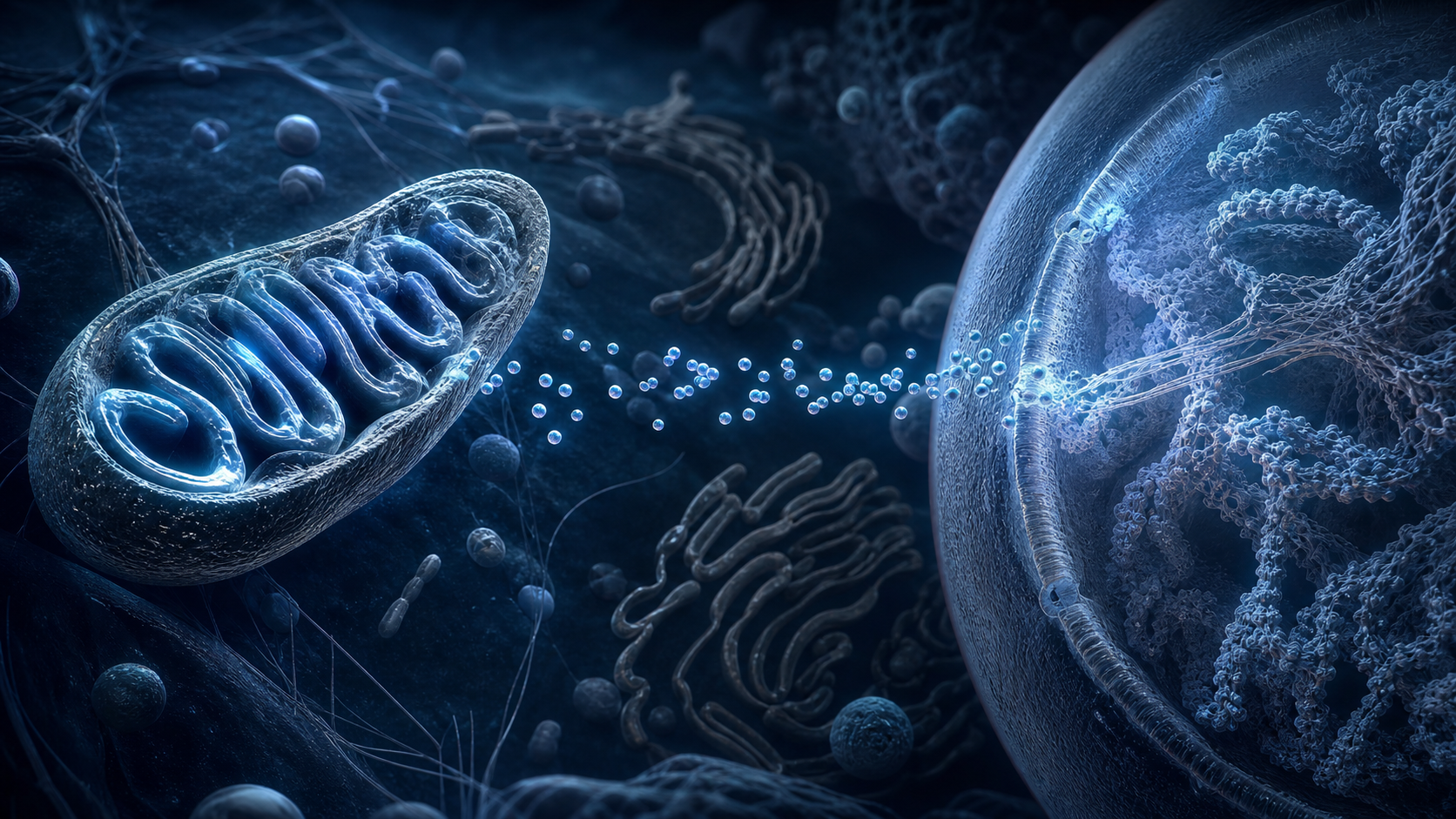
Colorful scientific visualization of mitochondrial cristae and respiratory complexes

Mitochondrial Peptide Overview

Inside the Mitochondria: What Makes SS-31 Interesting

Seven straightforward reasons SS-31, also called Elamipretide, remains important in mitochondrial and cellular-energy research.

Compound overview • 4 minute read

Quick Take

SS-31 is a mitochondria-targeted tetrapeptide that interacts with cardiolipin in the inner mitochondrial membrane. Researchers study whether that interaction can support cristae structure, electron transport, ATP production, oxidative balance, and tissue function under stress.

Why It Gets Attention

Most mitochondrial compounds act indirectly; SS-31 is designed to concentrate at the inner mitochondrial membrane. Its cardiolipin interaction gives researchers a direct structural link to energy production.

Studies can follow ATP output, oxygen consumption, reactive oxygen species, membrane potential, cristae structure, muscle performance, cardiac function, and kidney injury.

7 Key Areas Worth Knowing

The clearest themes are summarized below.

01

ATP Production

SS-31 research has reported improved mitochondrial energetic capacity and ATP production. ATP output, respiratory efficiency, and recovery after energy stress are central endpoints.

02

Cardiolipin and Cristae Protection

Cardiolipin helps organize the folded inner membrane where oxidative phosphorylation occurs. SS-31 studies examine cristae integrity, membrane structure, and cardiolipin oxidation.

03

Oxidative-Stress Control

Damaged mitochondria can produce excessive reactive oxygen species. Researchers track ROS, antioxidant capacity, lipid peroxidation, and oxidative damage.

04

Muscle Energy and Performance

Skeletal muscle depends on rapid mitochondrial ATP production. Human and animal studies have measured ATPmax, fatigue resistance, force, and mitochondrial ADP sensitivity.

05

Cardiac-Energy Research

The heart has constant energy demands and a dense mitochondrial network. SS-31 models examine cardiac output, systolic function, ischemic stress, and mitochondrial recovery.

06

Kidney Protection Models

Kidney cells also require large amounts of mitochondrial energy. Research has examined ischemic injury, diabetic kidney stress, fibrosis, apoptosis, and restoration of tubular function.

07

Healthy-Aging and Mitochondrial Resilience

Age-related decline is often associated with impaired mitochondrial responsiveness. SS-31 research tracks energetic capacity, muscle function, oxidative stress, and recovery in older models.

Why SS-31 Stands Out

It Targets the Inner Mitochondrial Membrane

SS-31 binds cardiolipin, a phospholipid concentrated in the inner mitochondrial membrane. That places the peptide directly beside the structures responsible for electron transport and ATP synthesis.

Structure and Energy Are Connected

Healthy cristae keep respiratory proteins organized. By studying cardiolipin, cristae shape, oxygen consumption, and ATP together, researchers can link membrane integrity with energy output.

Human ATP Measurements Add Translational Interest

A randomized study in older adults reported an acute increase in skeletal-muscle mitochondrial energetic capacity. That provides a direct human measurement rather than relying only on cell models.

The Same Mechanism Applies Across Tissues

Heart, skeletal muscle, and kidney cells all depend heavily on mitochondria. This shared biology explains why SS-31 appears across several organ and aging-related research models.

What Can Be Measured

These highlighted areas are most useful when treated as connected but separate endpoints. A controlled comparison can track atp production, cardiolipin and cristae protection, oxidative-stress control, muscle energy and performance, cardiac-energy research, kidney protection models, and healthy-aging and mitochondrial resilience at planned time points. This turns a broad question into clear observations and shows which part of the compound’s profile changes most strongly.

A strong design also connects each outcome with the biology behind it. Measurements can be paired with markers related to it targets the inner mitochondrial membrane, structure and energy are connected, human atp measurements add translational interest, and the same mechanism applies across tissues. Combining visible or functional results with mechanism-focused data makes the positive findings easier to interpret, repeat, and compare across the available cell, animal, and clinical literature.

Evidence and Limitations

SS-31 and Elamipretide studies use specific experimental or clinical-development formulations. Findings vary by model and endpoint and do not establish the performance of a separate research-use vial.

The Bottom Line

SS-31 stands out because its research story begins at the physical center of mitochondrial energy production. ATP, cardiolipin, oxidative stress, muscle performance, cardiac energy, kidney protection, and healthy aging all connect through inner-membrane function.

Sources

  1. SS-31, cardiolipin, cristae protection, and ATP recovery.
  2. Randomized Elamipretide study of muscle ATP production.
  3. SS-31 and oxidative stress in a diabetic kidney model.

Related Resources

Review SS-31 50mg or Open the Research Protocol

Research-use disclaimer: This article is for laboratory research education only. It does not provide medical advice or establish safety, efficacy, or suitability. Products discussed are not intended for human consumption, therapeutic use, diagnostic use, or veterinary use.

Posted on Leave a comment

Pinealon and the Architecture of Neural Aging

Silver-haired woman smiling while sketching botanical studies in a bright studio

Compound Spotlight

Pinealon and the Architecture of Neural Aging

A concise look at why the three-amino-acid peptide EDR keeps appearing in oxidative-stress, neuronal-structure, and cellular-aging studies.

Quick Take

Pinealon—also called EDR or Glu-Asp-Arg—is an experimental tripeptide studied mainly in cell and animal models. Its most interesting signals involve oxidative stress, neuronal viability, and preservation of dendritic structure. The evidence is early and does not establish clinical benefit.

What Pinealon Is

Pinealon is the common name used for EDR, a short chain made from glutamic acid, aspartic acid, and arginine. It belongs to a class of ultra-short peptides studied for possible effects on cellular signaling and stress-response pathways.

Its compact structure is part of the appeal. Instead of acting like a large protein, EDR is investigated as a small regulatory signal. Proposed mechanisms include changes in oxidative-stress handling, ERK-pathway activity, and gene expression, although the direct molecular explanation remains unsettled.

Four Reasons It Gets Attention

01

Oxidative-Stress Response

A 2011 cellular study associated Pinealon with lower reactive-oxygen-species accumulation and reduced necrotic cell death under experimentally induced stress.

02

Dendritic-Spine Preservation

In a mouse hippocampal-neuron model of amyloid toxicity, EDR restored the measured number of mature mushroom-shaped dendritic spines toward control levels.

03

Neural-Aging Models

A 2024 study using neurons derived from older human donors reported greater dendritic arborization and lower oxidative DNA damage after EDR exposure.

04

An Unusually Small Structure

At only three amino acids, EDR is used to explore how ultra-short peptides may influence signaling, gene expression, and cellular stress responses.

Blue-grey scientific visualization of a neuron with branching dendrites and dendritic spines
Conceptual visualization of neural branching and dendritic-spine structure.

What Individual Studies Found

Cellular stress: A 2011 study exposed several cell types to experimentally induced oxidative stress. Pinealon was associated with lower reactive-oxygen-species accumulation and reduced necrotic cell death, alongside changes in ERK signaling and cell-cycle activity.

Synaptic structure: In 2017, EDR increased mature mushroom-shaped dendritic spines in cultured mouse hippocampal neurons exposed to amyloid toxicity. A later 5xFAD mouse study also reported preservation of selected spine-density measures, although the response varied by sex and not every spine measure improved.

Cellular aging: A 2024 model converted fibroblasts from older donors into induced cortical neurons. EDR exposure was associated with greater dendritic branching and lower oxidative DNA damage. The same study did not find broad improvements across every aging marker, including mitochondrial, lysosomal, and p16 measurements.

What the Evidence Actually Supports

The clearest Pinealon findings are preclinical. Cell cultures and animal models provide useful signals, but they cannot establish effects in people, a validated route of administration, or a clinically effective dose.

The responsible conclusion is narrow: Pinealon is a legitimate experimental compound with recurring neurobiology themes. Human efficacy, pharmacokinetics, and long-term safety remain insufficiently defined, and the published work should not be interpreted as proof of a treatment outcome.

Sources

  1. Khavinson et al. Pinealon, reactive oxygen species, and cellular viability (2011).
  2. Kraskovskaya et al. EDR and neuronal spines in an in-vitro Alzheimer’s model (2017).
  3. Khavinson et al. EDR in a 5xFAD mouse model (2021).
  4. Ilina et al. Short peptides in induced neurons derived from older donors (2024).

Research-use disclaimer: This article is educational and does not provide medical advice or establish safety, efficacy, or suitability. Products discussed are for laboratory research use only and are not intended for human consumption.

Posted on Leave a comment

Tirzepatide: Why Dual-Agonist Signaling Matters

Scientific visualization of two receptor pathways converging inside a metabolic cell

Metabolic Peptide Overview

Tirzepatide: Why Dual-Agonist Signaling Matters

Seven straightforward reasons the combined GIP and GLP-1 receptor model remains central to modern metabolic research.

Compound overview • 4 minute read

Quick Take

Tirzepatide is a dual GIP and GLP-1 receptor agonist. Its two-receptor design gives researchers a practical model for studying appetite, glucose-dependent insulin release, insulin sensitivity, body weight, fat distribution, liver fat, and cardiometabolic markers.

Why It Gets Attention

Tirzepatide stands between GLP-1-only and triple-agonist research. It keeps the established GLP-1 pathway while adding GIP receptor activity, allowing researchers to examine whether the combined incretin signal changes metabolic outcomes.

Large randomized trials have reported clear changes in body weight and glucose regulation, while imaging and biomarker studies add information about liver fat, abdominal fat, insulin sensitivity, and beta-cell function.

7 Key Areas Worth Knowing

The clearest themes are summarized below.

01

Body-Weight Reduction

Randomized trials reported substantial and sustained reductions in body weight. Researchers can follow total change, percentage change, waist circumference, and the proportion reaching defined targets.

02

Glucose and HbA1c Control

The dual incretin signal supports glucose-dependent insulin activity. HbA1c, fasting glucose, post-meal glucose, and continuous-monitoring outcomes are common endpoints.

03

Appetite and Satiety

GLP-1 and GIP pathways influence how the body responds to food. Researchers examine hunger, fullness, food intake, cravings, and meal-related hormone signaling.

04

Insulin Sensitivity

Tirzepatide trials have reported improvements in insulin-sensitivity markers. This supports research involving fasting insulin, HOMA-IR, adiponectin, and glucose disposal.

05

Beta-Cell Function

Beta cells control insulin release in response to glucose. Research has tracked proinsulin, C-peptide, insulin secretion, and model-based beta-cell function.

06

Liver and Abdominal Fat

MRI research has reported reductions in liver fat and abdominal adipose tissue. These measurements add a clearer picture of metabolic change than body weight alone.

07

Cardiometabolic Markers

Weight and glucose changes often occur alongside broader metabolic shifts. Blood pressure, triglycerides, cholesterol, waist circumference, and inflammatory markers can all be followed.

Why Tirzepatide Stands Out

A Dual-Incretin Design

Tirzepatide activates both GIP and GLP-1 receptors. That makes it more mechanistically complete than a GLP-1-only model while remaining easier to interpret than a triple agonist.

Strong Weight and Glucose Data

SURMOUNT-1 reported large weight changes across studied doses. SURPASS-2 also found strong HbA1c and weight responses in a head-to-head comparison with Semaglutide.

Insulin Sensitivity and Beta Cells

Tirzepatide research goes beyond scale weight. Biomarker analyses have reported improvements in insulin sensitivity, beta-cell function, fasting proinsulin, and related measures.

Imaging Adds More Detail

The SURPASS-3 MRI substudy measured liver and abdominal fat directly. That provides researchers with objective body-composition and organ-fat endpoints.

What Can Be Measured

These highlighted areas are most useful when treated as connected but separate endpoints. A controlled comparison can track body-weight reduction, glucose and hba1c control, appetite and satiety, insulin sensitivity, beta-cell function, liver and abdominal fat, and cardiometabolic markers at planned time points. This turns a broad question into clear observations and shows which part of the compound’s profile changes most strongly.

A strong design also connects each outcome with the biology behind it. Measurements can be paired with markers related to a dual-incretin design, strong weight and glucose data, insulin sensitivity and beta cells, and imaging adds more detail. Combining visible or functional results with mechanism-focused data makes the positive findings easier to interpret, repeat, and compare across the available cell, animal, and clinical literature.

Evidence and Limitations

Published clinical results use regulated pharmaceutical formulations and treatment protocols. They do not automatically validate the identity, purity, or performance of a separate research-use material.

The Bottom Line

Tirzepatide remains one of the strongest dual-agonist research topics because its benefits are measurable across several systems. Weight, glucose, appetite, insulin sensitivity, beta-cell function, liver fat, and cardiometabolic markers all fit within one clear GIP/GLP-1 framework.

Sources

  1. SURMOUNT-1 Tirzepatide obesity trial.
  2. SURPASS-2 Tirzepatide versus Semaglutide trial.
  3. SURPASS-3 MRI liver and abdominal-fat substudy.

Related Resources

Review Tirzepatide 20mg or Open the Research Protocol

Research-use disclaimer: This article is for laboratory research education only. It does not provide medical advice or establish safety, efficacy, or suitability. Products discussed are not intended for human consumption, therapeutic use, diagnostic use, or veterinary use.

Posted on Leave a comment

Inside GLOW Blend: Three Peptides, One Skin-Focused Strategy

Active woman standing with a bicycle on a modern waterfront

Skin and Repair Blend Overview

Inside GLOW Blend: Three Peptides, One Skin-Focused Strategy

Six straightforward reasons the GHK-Cu, TB-500, and BPC-157 combination creates an engaging skin, collagen, and tissue-repair research model.

Compound overview • 4 minute read

Quick Take

GLOW combines 35mg GHK-Cu, 10mg TB-500, and 5mg BPC-157 in one research vial. The blend brings together collagen production, extracellular-matrix remodeling, cell migration, angiogenesis, inflammatory balance, wound closure, and broader connective-tissue research.

Why It Gets Attention

GLOW has a simple visual research story: tissue has to rebuild with the right cells, circulation, and structural proteins. GHK-Cu supplies the strongest collagen and matrix theme, while thymosin beta-4-related and BPC-157 research add cell-migration and local repair questions.

Researchers can follow collagen expression, fibroblast activity, wound closure, epithelial coverage, vessel density, inflammatory markers, oxidative stress, tissue strength, skin architecture, and restoration of normal connective-tissue organization.

6 Key Areas Worth Knowing

The clearest themes are summarized below.

01

Collagen Production

GHK-Cu research has repeatedly focused on stimulation of collagen synthesis and fibroblast activity. Type-specific collagen, procollagen markers, matrix density, and tissue organization can be measured.

02

Skin Structure and Elasticity

Collagen, elastin, and glycosaminoglycans help determine visible tissue architecture. Dermal thickness, elasticity markers, matrix quality, and age-related structural change are useful endpoints.

03

Wound Closure

All three components have appeared in tissue-repair or wound-related research. Closure rate, re-epithelialization, granulation tissue, collagen deposition, and tissue strength can be followed.

04

Cell Migration

Thymosin beta-4-related biology emphasizes actin and movement of repair-associated cells. Epithelial migration, endothelial movement, myoblast recruitment, and coverage of damaged areas are measurable.

05

Angiogenesis

New blood vessels support rebuilding by delivering oxygen and nutrients. Vessel density, endothelial activity, VEGF-related signaling, and stabilization of vascular networks are useful endpoints.

06

Inflammatory Balance

Repair depends on an organized inflammatory response that can transition into remodeling. Cytokines, immune-cell activity, swelling, oxidative stress, and resolution markers can be compared.

Why the GLOW Blend Stands Out

GHK-Cu Leads the Collagen Story

GHK-Cu is the largest component in the blend and gives GLOW a clear matrix-remodeling center. Fibroblasts, collagen expression, extracellular-matrix organization, and antioxidant signaling provide direct laboratory endpoints.

TB-500 Adds Cell Movement

Repair cells need to reach the damaged area before collagen can be organized. Thymosin beta-4-related actin and migration research adds an early-stage repair mechanism to the blend.

BPC-157 Adds Local Tissue Response

BPC-157 research connects with collagen organization, angiogenesis, and tissue strength in preclinical models. Those endpoints complement GHK-Cu and TB-500 without simply repeating their research roles.

Visible and Molecular Outcomes Can Be Paired

Skin and wound models allow researchers to compare appearance with tissue biology. Closure, thickness, elasticity, and structure can be measured alongside collagen genes, vessel markers, and inflammatory pathways.

What Can Be Measured

These highlighted areas are most useful when treated as connected but separate endpoints. A controlled comparison can track collagen production, skin structure and elasticity, wound closure, cell migration, angiogenesis, and inflammatory balance at planned time points. This turns a broad question into clear observations and shows which part of the compound’s profile changes most strongly.

A strong design also connects each outcome with the biology behind it. Measurements can be paired with markers related to ghk-cu leads the collagen story, tb-500 adds cell movement, bpc-157 adds local tissue response, and visible and molecular outcomes can be paired. Combining visible or functional results with mechanism-focused data makes the positive findings easier to interpret, repeat, and compare across the available cell, animal, and clinical literature.

Evidence and Limitations

The exact three-component GLOW Blend has not been evaluated in controlled human trials. Individual-component findings cannot establish the combined material’s interactions, safety, or performance.

The Bottom Line

GLOW stands out because its research themes move naturally from cell migration to collagen-rich tissue remodeling. Wound closure, skin structure, angiogenesis, inflammatory balance, and connective-tissue organization create an immediately understandable research profile.

Sources

  1. BPC-157 and tendon-healing research.
  2. Copper-peptide tissue-remodeling research.
  3. Thymosin beta-4 and wound-healing research.

Related Resources

Review GLOW Blend or Open the Research Protocol

Research-use disclaimer: This article is for laboratory research education only. It does not provide medical advice or establish safety, efficacy, or suitability. Products discussed are not intended for human consumption, therapeutic use, diagnostic use, or veterinary use.
Posted on Leave a comment

Semax, BDNF, and the Search for Neural Resilience

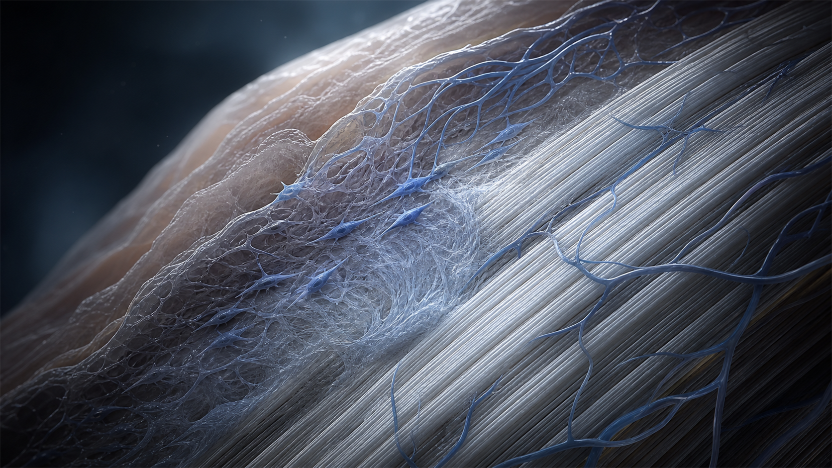
Scientific visualization of synapses and dendritic spine remodelling

Neurotrophic Peptide Overview

Semax, BDNF, and the Search for Neural Resilience

Six straightforward reasons Semax remains a recognizable peptide in BDNF, neuroprotection, learning, memory, and ischemia research.

Compound overview • 4 minute read

Quick Take

Semax is a synthetic heptapeptide derived from the ACTH(4-7) sequence with a Pro-Gly-Pro extension. Researchers study its relationship with BDNF, TrkB, neurotrophin gene expression, learning, memory, ischemic stress, and functional neurological recovery.

Why It Gets Attention

Semax has a strong research identity because it connects a short peptide with measurable neurotrophic signaling. Rat studies have reported changes in BDNF protein, BDNF transcripts, TrkB activation, and expression of several neurotrophins after cerebral ischemia.

Those molecular findings can be paired with conditioned learning, memory, neurological function, motor recovery, infarct measurements, oxidative stress, and transcriptional response to build a broad but coherent neuroscience model.

6 Key Areas Worth Knowing

The clearest themes are summarized below.

01

BDNF Signaling

Semax research has reported increased BDNF protein and gene expression in brain-related models. BDNF concentration, transcript levels, regional expression, and timing of the response are central endpoints.

02

TrkB Receptor Activation

BDNF becomes biologically meaningful through its TrkB receptor. Semax studies have measured TrkB phosphorylation and transcript changes, connecting neurotrophin production with receptor activity.

03

Learning and Conditioned Response

Animal studies have associated Semax with improved performance in conditioned-learning tasks. Acquisition speed, correct responses, retention, errors, and adaptability can all be measured.

04

Memory and Cognitive Models

Semax appears frequently in research involving hippocampal function and cognitive performance. Short-term memory, long-term retention, attention, object recognition, and task performance are useful endpoints.

05

Ischemic-Stress Research

A large part of Semax research examines brain response after experimental ischemia. Infarct size, neurological deficits, inflammatory markers, oxidative stress, and neuronal survival can be followed.

06

Functional-Recovery Measures

Human rehabilitation research has paired Semax with BDNF and functional outcome measurements. Motor scales, Barthel scores, rehabilitation timing, and recovery trajectories provide practical comparisons.

Why Semax Stands Out

BDNF and TrkB Form a Clear Mechanism

A rat hippocampus study reported increased BDNF, TrkB transcripts, and TrkB phosphorylation. That gives researchers a direct molecular path connecting peptide exposure with plasticity-related signaling.

The Neurotrophin Response Is Broader Than BDNF

Ischemia studies have reported time-dependent changes in NGF, NT-3, and several Trk receptors. This makes timing and brain region important parts of the Semax research design.

Molecular and Functional Outcomes Can Be Paired

BDNF levels alone do not show whether behavior or recovery changes. Combining molecular measurements with learning tasks, motor scales, and neurological outcomes creates a more complete picture.

Ischemia Provides a Structured Stress Model

Experimental cerebral ischemia produces defined molecular, structural, and functional changes. Researchers can use that model to compare neurotrophin expression, tissue damage, inflammation, and recovery over time.

What Can Be Measured

These highlighted areas are most useful when treated as connected but separate endpoints. A controlled comparison can track bdnf signaling, trkb receptor activation, learning and conditioned response, memory and cognitive models, ischemic-stress research, and functional-recovery measures at planned time points. This turns a broad question into clear observations and shows which part of the compound’s profile changes most strongly.

A strong design also connects each outcome with the biology behind it. Measurements can be paired with markers related to bdnf and trkb form a clear mechanism, the neurotrophin response is broader than bdnf, molecular and functional outcomes can be paired, and ischemia provides a structured stress model. Combining visible or functional results with mechanism-focused data makes the positive findings easier to interpret, repeat, and compare across the available cell, animal, and clinical literature.

Evidence and Limitations

Semax evidence includes animal studies and regionally published human research, with limited large independent trials. Findings should remain tied to the exact formulation, administration method, and research model.

The Bottom Line

Semax stands out because its neuroscience story connects molecular signaling with visible function. BDNF, TrkB, learning, memory, ischemic stress, neurotrophin transcription, and recovery measures can all be studied within one organized framework.

Sources

  1. Semax regulation of BDNF and TrkB in rat hippocampus.
  2. Semax and neurotrophin transcription after cerebral ischemia.
  3. Semax, BDNF, and functional recovery after ischemic stroke.

Related Resources

Review Semax 10mg or Open the Research Protocol

Research-use disclaimer: This article is for laboratory research education only. It does not provide medical advice or establish safety, efficacy, or suitability. Products discussed are not intended for human consumption, therapeutic use, diagnostic use, or veterinary use.